5J67 | pdb_00005j67

Structure of Astrotactin-2, a conserved vertebrate-specific and perforin-like membrane protein involved in neuronal development


Experimental Data Snapshot

  • Method: X-RAY DIFFRACTION
  • Resolution: 3.16 Å
  • R-Value Free: 
    0.259 (Depositor), 0.263 (DCC) 
  • R-Value Work: 
    0.233 (Depositor), 0.235 (DCC) 
  • R-Value Observed: 
    0.235 (Depositor) 

wwPDB Validation 3D Report Full Report

Validation slider image for 5J67

Ligand Structure Quality Assessment 


This is version 2.1 of the entry. See complete history

Literature

Structure of astrotactin-2: a conserved vertebrate-specific and perforin-like membrane protein involved in neuronal development.

Ni, T.Harlos, K.Gilbert, R.

(2016) Open Biol 6

  • DOI: https://doi.org/10.1098/rsob.160053
  • Primary Citation Related Structures: 
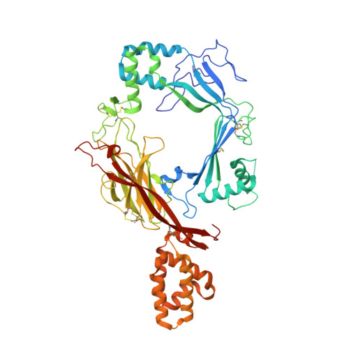
    5J67, 5J68, 5J69

  • PubMed Abstract: 

    The vertebrate-specific proteins astrotactin-1 and 2 (ASTN-1 and ASTN-2) are integral membrane perforin-like proteins known to play critical roles in neurodevelopment, while ASTN-2 has been linked to the planar cell polarity pathway in hair cells. Genetic variations associated with them are linked to a variety of neurodevelopmental disorders and other neurological pathologies, including an advanced onset of Alzheimer's disease. Here we present the structure of the majority endosomal region of ASTN-2, showing it to consist of a unique combination of polypeptide folds: a perforin-like domain, a minimal epidermal growth factor-like module, a unique form of fibronectin type III domain and an annexin-like domain. The perforin-like domain differs from that of other members of the membrane attack complex-perforin (MACPF) protein family in ways that suggest ASTN-2 does not form pores. Structural and biophysical data show that ASTN-2 (but not ASTN-1) binds inositol triphosphates, suggesting a mechanism for membrane recognition or secondary messenger regulation of its activity. The annexin-like domain is closest in fold to repeat three of human annexin V and similarly binds calcium, and yet shares no sequence homology with it. Overall, our structure provides the first atomic-resolution description of a MACPF protein involved in development, while highlighting distinctive features of ASTN-2 responsible for its activity.


  • Organizational Affiliation
    • Division of Structural Biology, Wellcome Trust Centre for Human Genetics, University of Oxford, Roosevelt Drive, Oxford OX3 7BN, UK.

Macromolecule Content 

  • Total Structure Weight: 269.67 kDa 
  • Atom Count: 18,450 
  • Modeled Residue Count: 2,237 
  • Deposited Residue Count: 2,288 
  • Unique protein chains: 1

Macromolecules

Find similar proteins by:|  3D Structure
Entity ID: 1
MoleculeChains  Sequence LengthOrganismDetailsImage
Astrotactin-2
A, B, C, D
572Homo sapiensMutation(s): 0 
Gene Names: ASTN2KIAA0634
UniProt & NIH Common Fund Data Resources
Find proteins for O75129 (Homo sapiens)
Explore O75129 
Go to UniProtKB:  O75129
PHAROS:  O75129
GTEx:  ENSG00000148219 
Entity Groups
Sequence Clusters30% Identity50% Identity70% Identity90% Identity95% Identity100% Identity
UniProt GroupO75129
Glycosylation
Glycosylation Sites: 2Go to GlyGen: O75129-1
Sequence Annotations
Expand
Reference Sequence

Oligosaccharides

Help  
Entity ID: 2
MoleculeChains Length2D Diagram GlycosylationD Interactions
alpha-D-mannopyranose-(1-2)-alpha-D-mannopyranose-(1-3)-[alpha-D-mannopyranose-(1-2)-alpha-D-mannopyranose-(1-6)]alpha-D-mannopyranose-(1-6)-[alpha-D-mannopyranose-(1-2)-alpha-D-mannopyranose-(1-3)]beta-D-mannopyranose-(1-4)-2-acetamido-2-deoxy-beta-D-glucopyranose-(1-4)-2-acetamido-2-deoxy-beta-D-glucopyranose
E, F, G, H
10N-Glycosylation
Glycosylation Resources
GlyTouCan: G89864BN
GlyCosmos: G89864BN
GlyGen: G89864BN

Small Molecules

Ligands 2 Unique
IDChains Name / Formula / InChI Key2D Diagram3D Interactions
I3P

Query on I3P


Download:Ideal Coordinates CCD File
J [auth A],
L [auth B],
N [auth C],
P [auth D]
D-MYO-INOSITOL-1,4,5-TRIPHOSPHATE
C6 H15 O15 P3
MMWCIQZXVOZEGG-XJTPDSDZSA-N
NAG

Query on NAG


Download:Ideal Coordinates CCD File
I [auth A],
K [auth B],
M [auth C],
O [auth D]
2-acetamido-2-deoxy-beta-D-glucopyranose
C8 H15 N O6
OVRNDRQMDRJTHS-FMDGEEDCSA-N

Experimental Data & Validation

Experimental Data

  • Method: X-RAY DIFFRACTION
  • Resolution: 3.16 Å
  • R-Value Free:  0.259 (Depositor), 0.263 (DCC) 
  • R-Value Work:  0.233 (Depositor), 0.235 (DCC) 
  • R-Value Observed: 0.235 (Depositor) 
Space Group: P 1
Unit Cell:
Length ( Å )Angle ( ˚ )
a = 101.24α = 87.18
b = 108.19β = 84.5
c = 111.36γ = 65.67
Software Package:
Software NamePurpose
REFMACrefinement
xia2data reduction
xia2data scaling
SHELXDEphasing
AutoSolphasing

Structure Validation

View Full Validation Report


Ligand Structure Quality Assessment 


Entry History 

& Funding Information

Deposition Data


Funding OrganizationLocationGrant Number
Medical Research Council (United Kingdom)United KingdomMR/N000331/1

Revision History  (Full details and data files)

  • Version 1.0: 2016-05-25
    Type: Initial release
  • Version 1.1: 2016-06-15
    Changes: Database references
  • Version 1.2: 2017-09-13
    Changes: Author supporting evidence
  • Version 1.3: 2019-04-03
    Changes: Data collection, Source and taxonomy
  • Version 2.0: 2020-07-29
    Type: Remediation
    Reason: Carbohydrate remediation
    Changes: Advisory, Atomic model, Data collection, Derived calculations, Source and taxonomy, Structure summary
  • Version 2.1: 2024-11-13
    Changes: Data collection, Database references, Derived calculations, Refinement description, Structure summary