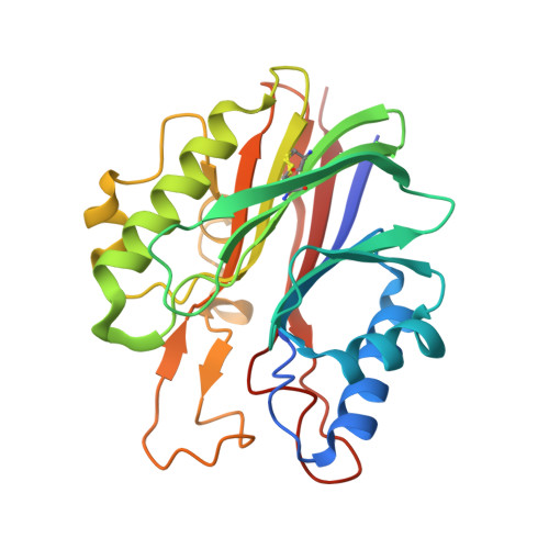
5J3Z

Crystal structure of m2hTDP2-CAT in complex with a small molecule inhibitor


Experimental Data Snapshot

  • Method: X-RAY DIFFRACTION
  • Resolution: 1.80 Å
  • R-Value Free: 0.202 
  • R-Value Work: 0.173 
  • R-Value Observed: 0.174 

Starting Model: experimental
View more details

wwPDB Validation   3D Report Full Report


Ligand Structure Quality Assessment 


This is version 1.3 of the entry. See complete history


Literature

Mode of action of DNA-competitive small molecule inhibitors of tyrosyl DNA phosphodiesterase 2.

Hornyak, P.Askwith, T.Walker, S.Komulainen, E.Paradowski, M.Pennicott, L.E.Bartlett, E.J.Brissett, N.C.Raoof, A.Watson, M.Jordan, A.M.Ogilvie, D.J.Ward, S.E.Atack, J.R.Pearl, L.H.Caldecott, K.W.Oliver, A.W.

(2016) Biochem J 473: 1869-1879

  • DOI: https://doi.org/10.1042/BCJ20160180
  • Primary Citation of Related Structures:  
    5J3P, 5J3S, 5J3Z, 5J42

  • PubMed Abstract: 

    Tyrosyl-DNA phosphodiesterase 2 (TDP2) is a 5'-tyrosyl DNA phosphodiesterase important for the repair of DNA adducts generated by non-productive (abortive) activity of topoisomerase II (TOP2). TDP2 facilitates therapeutic resistance to topoisomerase poisons, which are widely used in the treatment of a range of cancer types. Consequently, TDP2 is an interesting target for the development of small molecule inhibitors that could restore sensitivity to topoisomerase-directed therapies. Previous studies identified a class of deazaflavin-based molecules that showed inhibitory activity against TDP2 at therapeutically useful concentrations, but their mode of action was uncertain. We have confirmed that the deazaflavin series inhibits TDP2 enzyme activity in a fluorescence-based assay, suitable for high-throughput screen (HTS)-screening. We have gone on to determine crystal structures of these compounds bound to a 'humanized' form of murine TDP2. The structures reveal their novel mode of action as competitive ligands for the binding site of an incoming DNA substrate, and point the way to generating novel and potent inhibitors of TDP2.


  • Organizational Affiliation

    Genome Damage and Stability Centre, School of Life Sciences, University of Sussex, Falmer BN1 9RQ, U.K. Cancer Research UK DNA Repair Enzymes Group, Genome Damage and Stability Centre, School of Life Sciences, University of Sussex, Falmer BN1 9RQ, U.K.


Macromolecules
Find similar proteins by:  (by identity cutoff)  |  3D Structure
Entity ID: 1
MoleculeChains Sequence LengthOrganismDetailsImage
Tyrosyl-DNA phosphodiesterase 2
A, B
256Mus musculusMutation(s): 4 
Gene Names: Tdp2Ttrap
EC: 3.1.4
UniProt
Find proteins for Q9JJX7 (Mus musculus)
Explore Q9JJX7 
Go to UniProtKB:  Q9JJX7
Entity Groups  
Sequence Clusters30% Identity50% Identity70% Identity90% Identity95% Identity100% Identity
UniProt GroupQ9JJX7
Sequence Annotations
Expand
  • Reference Sequence
Small Molecules
Ligands 5 Unique
IDChains Name / Formula / InChI Key2D Diagram3D Interactions
6FQ
Query on 6FQ

Download Ideal Coordinates CCD File 
C [auth A],
S [auth B]
2,4-dioxo-10-[3-(1H-tetrazol-5-yl)phenyl]-2,3,4,10-tetrahydropyrimido[4,5-b]quinoline-8-carbonitrile
C19 H10 N8 O2
NDVGJTJBERLSGL-UHFFFAOYSA-N
GOL
Query on GOL

Download Ideal Coordinates CCD File 
E [auth A],
F [auth A],
U [auth B],
V [auth B]
GLYCEROL
C3 H8 O3
PEDCQBHIVMGVHV-UHFFFAOYSA-N
EDO
Query on EDO

Download Ideal Coordinates CCD File 
G [auth A]
H [auth A]
I [auth A]
J [auth A]
K [auth A]
G [auth A],
H [auth A],
I [auth A],
J [auth A],
K [auth A],
L [auth A],
M [auth A],
N [auth A],
O [auth A],
P [auth A],
Q [auth A],
W [auth B],
X [auth B],
Y [auth B],
Z [auth B]
1,2-ETHANEDIOL
C2 H6 O2
LYCAIKOWRPUZTN-UHFFFAOYSA-N
ACT
Query on ACT

Download Ideal Coordinates CCD File 
AA [auth B]
BA [auth B]
CA [auth B]
DA [auth B]
EA [auth B]
AA [auth B],
BA [auth B],
CA [auth B],
DA [auth B],
EA [auth B],
R [auth A]
ACETATE ION
C2 H3 O2
QTBSBXVTEAMEQO-UHFFFAOYSA-M
MG
Query on MG

Download Ideal Coordinates CCD File 
D [auth A],
T [auth B]
MAGNESIUM ION
Mg
JLVVSXFLKOJNIY-UHFFFAOYSA-N
Experimental Data & Validation

Experimental Data

  • Method: X-RAY DIFFRACTION
  • Resolution: 1.80 Å
  • R-Value Free: 0.202 
  • R-Value Work: 0.173 
  • R-Value Observed: 0.174 
  • Space Group: P 1 21 1
Unit Cell:
Length ( Å )Angle ( ˚ )
a = 61.01α = 90
b = 42.8β = 94.15
c = 108.76γ = 90
Software Package:
Software NamePurpose
BUSTERrefinement
XDSdata reduction
Aimlessdata scaling
PHASERphasing

Structure Validation

View Full Validation Report


Ligand Structure Quality Assessment 


Entry History & Funding Information

Deposition Data


Funding OrganizationLocationGrant Number
Cancer Research UKUnited KingdomC302/A14532
Cancer Research UKUnited KingdomC6563/A16771
Cancer Research UKUnited KingdomC480/A11411

Revision History  (Full details and data files)

  • Version 1.0: 2016-05-04
    Type: Initial release
  • Version 1.1: 2016-07-06
    Changes: Database references
  • Version 1.2: 2024-01-10
    Changes: Data collection, Database references, Derived calculations, Refinement description
  • Version 1.3: 2024-10-09
    Changes: Structure summary