5J1E | pdb_00005j1e

Crystal Structure of a Hydroxypyridone Carboxylic Acid Active-Site RNase H Inhibitor in Complex with HIV Reverse Transcriptase


Experimental Data Snapshot

  • Method: X-RAY DIFFRACTION
  • Resolution: 2.90 Å
  • R-Value Free: 
    0.294 (Depositor), 0.299 (DCC) 
  • R-Value Work: 
    0.254 (Depositor), 0.259 (DCC) 
  • R-Value Observed: 
    0.256 (Depositor) 

Starting Model: experimental
View more details

wwPDB Validation 3D Report Full Report

Validation slider image for 5J1E

Ligand Structure Quality Assessment 


This is version 1.4 of the entry. See complete history

Literature

Design, Synthesis, and Biological Evaluations of Hydroxypyridonecarboxylic Acids as Inhibitors of HIV Reverse Transcriptase Associated RNase H.

Kankanala, J.Kirby, K.A.Liu, F.Miller, L.Nagy, E.Wilson, D.J.Parniak, M.A.Sarafianos, S.G.Wang, Z.

(2016) J Med Chem 59: 5051-5062

  • DOI: https://doi.org/10.1021/acs.jmedchem.6b00465
  • Primary Citation Related Structures: 
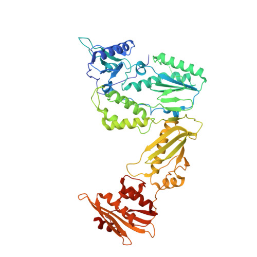
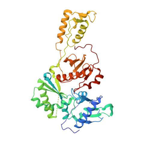
    5J1E

  • PubMed Abstract: 

    Targeting the clinically unvalidated reverse transcriptase (RT) associated ribonuclease H (RNase H) for human immunodeficiency virus (HIV) drug discovery generally entails chemotypes capable of chelating two divalent metal ions in the RNase H active site. The hydroxypyridonecarboxylic acid scaffold has been implicated in inhibiting homologous HIV integrase (IN) and influenza endonuclease via metal chelation. We report herein the design, synthesis, and biological evaluations of a novel variant of the hydroxypyridonecarboxylic acid scaffold featuring a crucial N-1 benzyl or biarylmethyl moiety. Biochemical studies show that most analogues consistently inhibited HIV RT-associated RNase H in the low micromolar range in the absence of significant inhibition of RT polymerase or IN. One compound showed reasonable cell-based antiviral activity (EC50 = 10 μM). Docking and crystallographic studies corroborate favorable binding to the active site of HIV RNase H, providing a basis for the design of more potent analogues.


  • Organizational Affiliation
    • Center for Drug Design, Academic Health Center, University of Minnesota , Minneapolis, Minnesota 55455, United States.

Macromolecule Content 

  • Total Structure Weight: 228.88 kDa 
  • Atom Count: 15,484 
  • Modeled Residue Count: 1,887 
  • Deposited Residue Count: 1,972 
  • Unique protein chains: 2

Macromolecules

Find similar proteins by:|  3D Structure
Entity ID: 1
MoleculeChains  Sequence LengthOrganismDetailsImage
HIV-1 REVERSE TRANSCRIPTASE P66 DOMAIN
A, C
557Human immunodeficiency virus type 1 BH10Mutation(s): 1 
Gene Names: gag-pol
EC: 2.7.7.49 (PDB Primary Data), 2.7.7.7 (PDB Primary Data), 3.1.26.4 (PDB Primary Data)
UniProt
Find proteins for P03366 (Human immunodeficiency virus type 1 group M subtype B (isolate BH10))
Explore P03366 
Go to UniProtKB:  P03366
Entity Groups
Sequence Clusters30% Identity50% Identity70% Identity90% Identity95% Identity100% Identity
UniProt GroupP03366
Sequence Annotations
Expand
Reference Sequence
Find similar proteins by:|  3D Structure
Entity ID: 2
MoleculeChains  Sequence LengthOrganismDetailsImage
HIV-1 REVERSE TRANSCRIPTASE P51 DOMAIN
B, D
429Human immunodeficiency virus type 1 BH10Mutation(s): 1 
Gene Names: gag-pol
EC: 2.7.7.49 (PDB Primary Data), 2.7.7.7 (PDB Primary Data), 3.1.26.4 (PDB Primary Data)
UniProt
Find proteins for P03366 (Human immunodeficiency virus type 1 group M subtype B (isolate BH10))
Explore P03366 
Go to UniProtKB:  P03366
Entity Groups
Sequence Clusters30% Identity50% Identity70% Identity90% Identity95% Identity100% Identity
UniProt GroupP03366
Sequence Annotations
Expand
Reference Sequence

Experimental Data & Validation

Experimental Data

  • Method: X-RAY DIFFRACTION
  • Resolution: 2.90 Å
  • R-Value Free:  0.294 (Depositor), 0.299 (DCC) 
  • R-Value Work:  0.254 (Depositor), 0.259 (DCC) 
  • R-Value Observed: 0.256 (Depositor) 
Space Group: P 1
Unit Cell:
Length ( Å )Angle ( ˚ )
a = 69.07α = 105.47
b = 89.31β = 92.69
c = 108.22γ = 110.8
Software Package:
Software NamePurpose
PHENIXrefinement
XSCALEdata scaling
PHASERphasing
PDB_EXTRACTdata extraction
XDSdata reduction

Structure Validation

View Full Validation Report


Ligand Structure Quality Assessment 


Entry History 

& Funding Information

Deposition Data


Funding OrganizationLocationGrant Number
National Institutes of Health/National Institute Of Allergy and Infectious Diseases (NIH/NIAID)United StatesAI100890

Revision History  (Full details and data files)

  • Version 1.0: 2016-06-15
    Type: Initial release
  • Version 1.1: 2016-06-29
    Changes: Database references
  • Version 1.2: 2017-09-20
    Changes: Author supporting evidence, Database references, Derived calculations
  • Version 1.3: 2019-12-11
    Changes: Author supporting evidence
  • Version 1.4: 2023-09-27
    Changes: Data collection, Database references, Refinement description