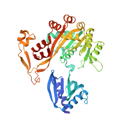
5IT5 | pdb_00005it5

Thermus thermophilus PilB core ATPase region


Experimental Data Snapshot

  • Method: X-RAY DIFFRACTION
  • Resolution: 2.65 Å
  • R-Value Free: 
    0.274 (Depositor), 0.280 (DCC) 
  • R-Value Work: 
    0.208 (Depositor), 0.210 (DCC) 
  • R-Value Observed: 
    0.210 (Depositor) 

Starting Model: experimental
View more details

wwPDB Validation   3D Report Full Report


Ligand Structure Quality Assessment 

Created with Raphaël 2.3.0Worse 01 BetterLigand structure goodness of fit to experimental dataBest fitted AGSClick on this verticalbar to view detailsBest fitted ATPClick on this verticalbar to view details

This is version 1.5 of the entry. See complete history


Literature

Crystal Structure of a Type IV Pilus Assembly ATPase: Insights into the Molecular Mechanism of PilB from Thermus thermophilus.

Mancl, J.M.Black, W.P.Robinson, H.Yang, Z.Schubot, F.D.

(2016) Structure 24: 1886-1897

  • DOI: https://doi.org/10.1016/j.str.2016.08.010
  • Primary Citation of Related Structures:  
    5IT5

  • PubMed Abstract: 

    Type IV pili (T4P) mediate bacterial motility and virulence. The PilB/GspE family ATPases power the assembly of T4P and type 2 secretion systems. We determined the structure of the ATPase region of PilB (PilB ATP ) in complex with ATPγS to provide a model of a T4P assembly ATPase and a view of a PilB/GspE family hexamer at better than 3-Å resolution. Spatial positioning and conformations of the protomers suggest a mechanism of force generation. All six PilB ATP protomers contain bound ATPγS. Two protomers form a closed conformation poised for ATP hydrolysis. The other four molecules assume an open conformation but separate into two pairs with distinct active-site accessibilities. We propose that one pair represents the post-hydrolysis phase while the other pair appears poised for ADP/ATP exchange. Collectively, the data suggest that T4P assembly is powered by coordinating concurrent substrate binding with ATP hydrolysis across the PilB hexamer.


  • Organizational Affiliation

    Department of Biological Sciences, Virginia Polytechnic Institute and State University, 125 Life Sciences 1 (MC 0910), 970 Washington Street Southwest, Blacksburg, VA 24061, USA.


Macromolecules
Find similar proteins by:  (by identity cutoff)  |  3D Structure
Entity ID: 1
MoleculeChains Sequence LengthOrganismDetailsImage
ATP binding motif-containing protein PilF385Thermus thermophilusMutation(s): 0 
Gene Names: pilF
UniProt
Find proteins for Q5SLC9 (Thermus thermophilus (strain ATCC 27634 / DSM 579 / HB8))
Explore Q5SLC9 
Go to UniProtKB:  Q5SLC9
Entity Groups  
Sequence Clusters30% Identity50% Identity70% Identity90% Identity95% Identity100% Identity
UniProt GroupQ5SLC9
Sequence Annotations
Expand
  • Reference Sequence
Small Molecules
Ligands 4 Unique
IDChains Name / Formula / InChI Key2D Diagram3D Interactions
AGS
Query on AGS

Download Ideal Coordinates CCD File 
I [auth A],
K [auth B],
N [auth E],
Q [auth C],
S [auth D]
PHOSPHOTHIOPHOSPHORIC ACID-ADENYLATE ESTER
C10 H16 N5 O12 P3 S
NLTUCYMLOPLUHL-KQYNXXCUSA-N
ATP
Query on ATP

Download Ideal Coordinates CCD File 
H [auth F]ADENOSINE-5'-TRIPHOSPHATE
C10 H16 N5 O13 P3
ZKHQWZAMYRWXGA-KQYNXXCUSA-N
ZN
Query on ZN

Download Ideal Coordinates CCD File 
G [auth F]
J [auth A]
L [auth B]
O [auth E]
R [auth C]
ZINC ION
Zn
PTFCDOFLOPIGGS-UHFFFAOYSA-N
MG
Query on MG

Download Ideal Coordinates CCD File 
M [auth B],
P [auth E]
MAGNESIUM ION
Mg
JLVVSXFLKOJNIY-UHFFFAOYSA-N
Experimental Data & Validation

Experimental Data

  • Method: X-RAY DIFFRACTION
  • Resolution: 2.65 Å
  • R-Value Free:  0.274 (Depositor), 0.280 (DCC) 
  • R-Value Work:  0.208 (Depositor), 0.210 (DCC) 
  • R-Value Observed: 0.210 (Depositor) 
Space Group: P 2 21 21
Unit Cell:
Length ( Å )Angle ( ˚ )
a = 107.55α = 90
b = 133.56β = 90
c = 208.42γ = 90
Software Package:
Software NamePurpose
PHENIXrefinement
iMOSFLMdata reduction
SCALAdata scaling
PHASERphasing

Structure Validation

View Full Validation Report


Ligand Structure Quality Assessment 

Created with Raphaël 2.3.0Worse 01 BetterLigand structure goodness of fit to experimental dataBest fitted AGSClick on this verticalbar to view detailsBest fitted ATPClick on this verticalbar to view details

Entry History & Funding Information

Deposition Data


Funding OrganizationLocationGrant Number
National Science Foundation (NSF, United States)United StatesMCB-1417726

Revision History  (Full details and data files)

  • Version 1.0: 2016-10-19
    Type: Initial release
  • Version 1.1: 2016-10-26
    Changes: Database references
  • Version 1.2: 2016-11-09
    Changes: Database references
  • Version 1.3: 2017-09-20
    Changes: Author supporting evidence, Database references, Derived calculations
  • Version 1.4: 2019-11-27
    Changes: Author supporting evidence
  • Version 1.5: 2023-09-27
    Changes: Data collection, Database references, Derived calculations, Refinement description