


Crystal structure of Esterase mutant - F72G
Seok, S.-H., Seo, M.-D., Kim, J., Ryu, Y.To be published.
Experimental Data Snapshot
wwPDB Validation 3D Report Full Report

Entity ID: 1 | |||||
|---|---|---|---|---|---|
| Molecule | Chains | Sequence Length | Organism | Details | Image |
| Esterase | 362 | uncultured bacterium | Mutation(s): 1 | 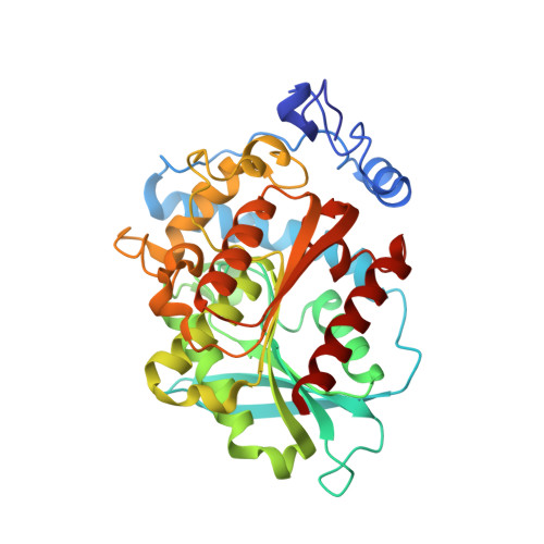 | |
| Length ( Å ) | Angle ( ˚ ) |
|---|---|
| a = 196.458 | α = 90 |
| b = 95.454 | β = 96.58 |
| c = 99.266 | γ = 90 |
| Software Name | Purpose |
|---|---|
| PHENIX | refinement |
| HKL-2000 | data processing |
| HKL-2000 | data scaling |
| PHENIX | phasing |


