5IJV | pdb_00005ijv

Crystal structure of bovine Fab E03


Experimental Data Snapshot

  • Method: X-RAY DIFFRACTION
  • Resolution: 2.20 Å
  • R-Value Free: 
    0.247 (Depositor), 0.248 (DCC) 
  • R-Value Work: 
    0.204 (Depositor), 0.207 (DCC) 
  • R-Value Observed: 
    0.206 (Depositor) 

Starting Model: experimental
View more details

wwPDB Validation 3D Report Full Report

Validation slider image for 5IJV

This is version 1.3 of the entry. See complete history

Literature

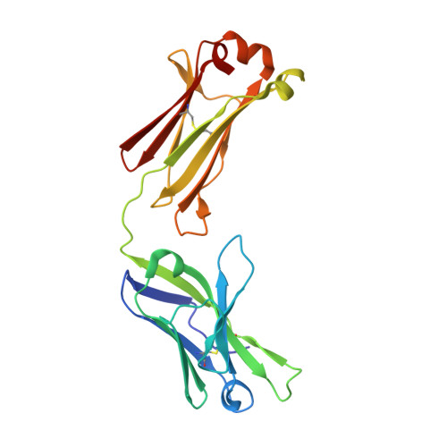
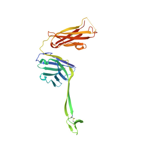
Conservation and diversity in the ultralong third heavy-chain complementarity-determining region of bovine antibodies.

Stanfield, R.L.Wilson, I.A.Smider, V.V.

(2016) Sci Immunol 1

  • DOI: https://doi.org/10.1126/sciimmunol.aaf7962
  • Primary Citation Related Structures: 
    5IHU, 5IJV, 5ILT

  • PubMed Abstract: 

    A subset of bovine antibodies have an exceptionally long third heavy-chain complementarity determining region (CDR H3) that is highly variable in sequence and includes multiple cysteines. These long CDR H3s (up to 69 residues) fold into a long stalk atop which sits a knob domain that is located far from the antibody surface. Three new bovine Fab crystal structures have been determined to decipher the conserved and variable features of ultralong CDR H3s that lead to diversity in antigen recognition. Despite high sequence variability, the stalks adopt a conserved β-ribbon structure, while the knob regions share a conserved β-sheet that serves as a scaffold for two connecting loops of variable length and conformation, as well as one conserved disulfide. Variation in patterns and connectivity of the remaining disulfides contribute to the knob structural diversity. The unusual architecture of these ultralong bovine CDR H3s for generating diversity is unique in adaptive immune systems.


  • Organizational Affiliation
    • Department of Integrative Structural and Computational Biology, The Scripps Research Institute, La Jolla, California, 92037, USA.

Macromolecule Content 

  • Total Structure Weight: 197.46 kDa 
  • Atom Count: 13,922 
  • Modeled Residue Count: 1,842 
  • Deposited Residue Count: 1,880 
  • Unique protein chains: 2

Macromolecules

Find similar proteins by:|  3D Structure
Entity ID: 1
MoleculeChains  Sequence LengthOrganismDetailsImage
bovine Fab E03 light chainA [auth L],
C [auth A],
E [auth C],
G [auth E]
216Bos taurusMutation(s): 0 
UniProt
Find proteins for Q3T101 (Bos taurus)
Explore Q3T101 
Go to UniProtKB:  Q3T101
Entity Groups
Sequence Clusters30% Identity50% Identity70% Identity90% Identity95% Identity100% Identity
UniProt GroupQ3T101
Sequence Annotations
Expand
Reference Sequence
Find similar proteins by:|  3D Structure
Entity ID: 2
MoleculeChains  Sequence LengthOrganismDetailsImage
bovine Fab E03 heavy chainB [auth H],
D [auth B],
F [auth D],
H [auth F]
254Bos taurusMutation(s): 0 

Experimental Data & Validation

Experimental Data

  • Method: X-RAY DIFFRACTION
  • Resolution: 2.20 Å
  • R-Value Free:  0.247 (Depositor), 0.248 (DCC) 
  • R-Value Work:  0.204 (Depositor), 0.207 (DCC) 
  • R-Value Observed: 0.206 (Depositor) 
Space Group: P 1 21 1
Unit Cell:
Length ( Å )Angle ( ˚ )
a = 62.244α = 90
b = 221.737β = 104.81
c = 67.947γ = 90
Software Package:
Software NamePurpose
PHENIXrefinement
DENZOdata reduction
SCALEPACKdata scaling
PHASERphasing

Structure Validation

View Full Validation Report


Entry History 

Deposition Data

Revision History  (Full details and data files)

  • Version 1.0: 2016-10-05
    Type: Initial release
  • Version 1.1: 2020-01-29
    Changes: Derived calculations
  • Version 1.2: 2023-09-27
    Changes: Data collection, Database references, Refinement description
  • Version 1.3: 2024-10-16
    Changes: Structure summary