5IJS | pdb_00005ijs

Crystal structure of autotaxin with orthovanadate bound as a trigonal bipyramidal intermediate analog


Experimental Data Snapshot

  • Method: X-RAY DIFFRACTION
  • Resolution: 2.20 Å
  • R-Value Free: 
    0.239 (Depositor), 0.239 (DCC) 
  • R-Value Work: 
    0.203 (Depositor), 0.209 (DCC) 
  • R-Value Observed: 
    0.204 (Depositor) 

Starting Model: experimental
View more details

wwPDB Validation 3D Report Full Report

Validation slider image for 5IJS

Ligand Structure Quality Assessment 


This is version 3.2 of the entry. See complete history

Literature

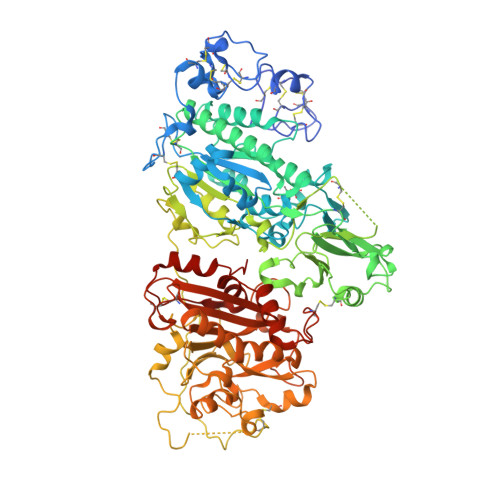
Structural snapshots of the catalytic cycle of the phosphodiesterase Autotaxin.

Hausmann, J.Keune, W.J.Hipgrave Ederveen, A.L.van Zeijl, L.Joosten, R.P.Perrakis, A.

(2016) J Struct Biol 195: 199-206

  • DOI: https://doi.org/10.1016/j.jsb.2016.06.002
  • Primary Citation Related Structures: 
    5IJQ, 5IJS

  • PubMed Abstract: 

    Autotaxin (ATX) is a secreted phosphodiesterase that produces the signalling lipid lysophosphatidic acid (LPA). The bimetallic active site of ATX is structurally related to the alkaline phosphatase superfamily. Here, we present a new crystal structure of ATX in complex with orthovanadate (ATX-VO5), which binds the Oγ nucleophile of Thr209 and adopts a trigonal bipyramidal conformation, following the nucleophile attack onto the substrate. We have now a portfolio of ATX structures we discuss as intermediates of the catalytic mechanism: the new ATX-VO5 structure; a unique structure where the nucleophile Thr209 is phosphorylated (ATX-pThr). Comparing these to a complex with the LPA product (ATX-LPA) and with a complex with a phosphate ion (ATX-PO4), that represent the Michaelis complex of the reaction, we observe movements of Thr209, changes in the relative displacement of the zinc ions, and a water molecule that likely fulfils the second nucleophilic attack. We propose that ATX follows the associative two-step in-line displacement mechanism.


  • Organizational Affiliation
    • Division of Biochemistry, The Netherlands Cancer Institute, Plesmanlaan 121, 1066 CX Amsterdam, The Netherlands. Electronic address: jens.hausmann@mpi-muenster.mpg.de.

Macromolecule Content 

  • Total Structure Weight: 96.96 kDa 
  • Atom Count: 6,559 
  • Modeled Residue Count: 784 
  • Deposited Residue Count: 827 
  • Unique protein chains: 1

Macromolecules

Find similar proteins by:|  3D Structure
Entity ID: 1
MoleculeChains  Sequence LengthOrganismDetailsImage
Ectonucleotide pyrophosphatase/phosphodiesterase family member 2827Rattus norvegicusMutation(s): 2 
Gene Names: Enpp2AtxNpps2
EC: 3.1.4.39 (PDB Primary Data), 3.1.4.4 (UniProt)
UniProt
Find proteins for Q64610 (Rattus norvegicus)
Explore Q64610 
Go to UniProtKB:  Q64610
Entity Groups
Sequence Clusters30% Identity50% Identity70% Identity90% Identity95% Identity100% Identity
UniProt GroupQ64610
Glycosylation
Glycosylation Sites: 1Go to GlyGen: Q64610-1
Sequence Annotations
Expand
Reference Sequence

Oligosaccharides

Help  
Entity ID: 2
MoleculeChains Length2D Diagram GlycosylationD Interactions
2-acetamido-2-deoxy-beta-D-glucopyranose-(1-4)-2-acetamido-2-deoxy-beta-D-glucopyranose
B
2N-Glycosylation
Glycosylation Resources
GlyTouCan: G42666HT
GlyCosmos: G42666HT
GlyGen: G42666HT

Small Molecules

Ligands 5 Unique
IDChains Name / Formula / InChI Key2D Diagram3D Interactions
5JK

Query on 5JK


Download:Ideal Coordinates CCD File
C [auth A]7alpha-hydroxycholesterol
C27 H46 O2
OYXZMSRRJOYLLO-RVOWOUOISA-N
IOD

Query on IOD


Download:Ideal Coordinates CCD File
G [auth A]
H [auth A]
I [auth A]
J [auth A]
K [auth A]
G [auth A],
H [auth A],
I [auth A],
J [auth A],
K [auth A],
L [auth A]
IODIDE ION
I
XMBWDFGMSWQBCA-UHFFFAOYSA-M
ZN

Query on ZN


Download:Ideal Coordinates CCD File
D [auth A],
E [auth A]
ZINC ION
Zn
PTFCDOFLOPIGGS-UHFFFAOYSA-N
CA

Query on CA


Download:Ideal Coordinates CCD File
F [auth A]CALCIUM ION
Ca
BHPQYMZQTOCNFJ-UHFFFAOYSA-N
NA

Query on NA


Download:Ideal Coordinates CCD File
M [auth A],
N [auth A]
SODIUM ION
Na
FKNQFGJONOIPTF-UHFFFAOYSA-N
Modified Residues  1 Unique
IDChains TypeFormula2D DiagramParent
6BR
Query on 6BR
A
L-PEPTIDE LINKINGC4 H8 N O7 VTHR

Experimental Data & Validation

Experimental Data

  • Method: X-RAY DIFFRACTION
  • Resolution: 2.20 Å
  • R-Value Free:  0.239 (Depositor), 0.239 (DCC) 
  • R-Value Work:  0.203 (Depositor), 0.209 (DCC) 
  • R-Value Observed: 0.204 (Depositor) 
Space Group: P 1
Unit Cell:
Length ( Å )Angle ( ˚ )
a = 53.75α = 99.33
b = 63.45β = 105.91
c = 70.55γ = 99.51
Software Package:
Software NamePurpose
MOSFLMdata reduction
SCALAdata scaling
PHASERphasing
REFMACrefinement
PDB_EXTRACTdata extraction
Cootmodel building
PDB-REDOrefinement

Structure Validation

View Full Validation Report


Ligand Structure Quality Assessment 


Entry History 

& Funding Information

Deposition Data


Funding OrganizationLocationGrant Number
KWFNetherlands--
NWONetherlands--
NWONetherlands723.013.003

Revision History  (Full details and data files)

  • Version 1.0: 2016-06-15
    Type: Initial release
  • Version 1.1: 2016-06-22
    Changes: Database references
  • Version 1.2: 2016-07-13
    Changes: Database references
  • Version 2.0: 2019-04-24
    Changes: Data collection, Polymer sequence
  • Version 3.0: 2020-07-29
    Type: Remediation
    Reason: Carbohydrate remediation
    Changes: Atomic model, Data collection, Derived calculations, Refinement description, Structure summary
  • Version 3.1: 2024-01-10
    Changes: Data collection, Database references, Derived calculations, Refinement description, Structure summary
  • Version 3.2: 2025-07-30
    Changes: Structure summary