5IIZ | pdb_00005iiz

Xanthomonas campestris Peroxiredoxin Q - Structure F0


Experimental Data Snapshot

  • Method: X-RAY DIFFRACTION
  • Resolution: 1.05 Å
  • R-Value Free: 
    0.153 (Depositor), 0.152 (DCC) 
  • R-Value Work: 
    0.135 (Depositor), 0.134 (DCC) 
  • R-Value Observed: 
    0.136 (Depositor) 

Starting Model: experimental
View more details

wwPDB Validation 3D Report Full Report

Validation slider image for 5IIZ

This is version 1.5 of the entry. See complete history

Literature

Peroxiredoxin Catalysis at Atomic Resolution.

Perkins, A.Parsonage, D.Nelson, K.J.Ogba, O.M.Cheong, P.H.Poole, L.B.Karplus, P.A.

(2016) Structure 24: 1668-1678

  • DOI: https://doi.org/10.1016/j.str.2016.07.012
  • Primary Citation Related Structures: 
    5IIZ, 5IM9, 5IMA, 5IMC, 5IMD, 5IMF, 5IMV, 5IMZ, 5INY, 5IO0, 5IO2, 5IOW, 5IOX, 5IPG, 5IPH

  • PubMed Abstract: 

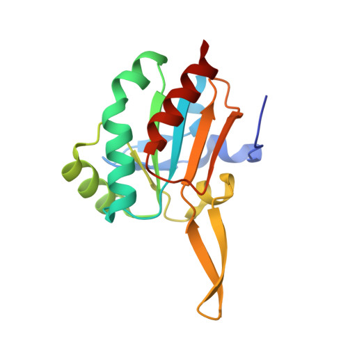
    Peroxiredoxins (Prxs) are ubiquitous cysteine-based peroxidases that guard cells against oxidative damage, are virulence factors for pathogens, and are involved in eukaryotic redox regulatory pathways. We have analyzed catalytically active crystals to capture atomic resolution snapshots of a PrxQ subfamily enzyme (from Xanthomonas campestris) proceeding through thiolate, sulfenate, and sulfinate species. These analyses provide structures of unprecedented accuracy for seeding theoretical studies, and reveal conformational intermediates giving insight into the reaction pathway. Based on a highly non-standard geometry seen for the sulfenate intermediate, we infer that the sulfenate formation itself can strongly promote local unfolding of the active site to enhance productive catalysis. Further, these structures reveal that preventing local unfolding, in this case via crystal contacts, results in facile hyperoxidative inactivation even for Prxs normally resistant to such inactivation. This supports previous proposals that conformation-specific inhibitors may be useful for achieving selective inhibition of Prxs that are drug targets.


  • Organizational Affiliation
    • Department of Biochemistry and Biophysics, Oregon State University, Corvallis, OR 97331, USA.

Macromolecule Content 

  • Total Structure Weight: 17.31 kDa 
  • Atom Count: 1,553 
  • Modeled Residue Count: 157 
  • Deposited Residue Count: 160 
  • Unique protein chains: 1

Macromolecules

Find similar proteins by:|  3D Structure
Entity ID: 1
MoleculeChains  Sequence LengthOrganismDetailsImage
Bacterioferritin comigratory protein160Xanthomonas campestris pv. raphani 756CMutation(s): 1 
Gene Names: bcpXCR_1978

Experimental Data & Validation

Experimental Data

  • Method: X-RAY DIFFRACTION
  • Resolution: 1.05 Å
  • R-Value Free:  0.153 (Depositor), 0.152 (DCC) 
  • R-Value Work:  0.135 (Depositor), 0.134 (DCC) 
  • R-Value Observed: 0.136 (Depositor) 
Space Group: P 1 21 1
Unit Cell:
Length ( Å )Angle ( ˚ )
a = 35.4α = 90
b = 51.36β = 103.27
c = 39.955γ = 90
Software Package:
Software NamePurpose
PHENIXrefinement
iMOSFLMdata reduction
SCALAdata scaling
PHASERphasing

Structure Validation

View Full Validation Report


Entry History 

& Funding Information

Deposition Data


Funding OrganizationLocationGrant Number
National Institutes of Health/National Institute of General Medical Sciences (NIH/NIGMS)United States--

Revision History  (Full details and data files)

  • Version 1.0: 2016-09-21
    Type: Initial release
  • Version 1.1: 2016-10-19
    Changes: Database references
  • Version 1.2: 2017-09-27
    Changes: Author supporting evidence, Database references, Derived calculations, Refinement description
  • Version 1.3: 2019-12-25
    Changes: Author supporting evidence
  • Version 1.4: 2023-09-27
    Changes: Data collection, Database references, Derived calculations, Refinement description
  • Version 1.5: 2024-10-09
    Changes: Structure summary