5IFO | pdb_00005ifo

X-ray structure of HSA-Myr-KP1019


Experimental Data Snapshot

  • Method: X-RAY DIFFRACTION
  • Resolution: 3.20 Å
  • R-Value Free: 
    0.262 (Depositor), 0.273 (DCC) 
  • R-Value Work: 
    0.244 (Depositor), 0.249 (DCC) 
  • R-Value Observed: 
    0.245 (Depositor) 

Starting Model: experimental
View more details

wwPDB Validation 3D Report Full Report

Validation slider image for 5IFO

Ligand Structure Quality Assessment 


This is version 1.6 of the entry. See complete history

Literature

X-ray Structure Analysis of Indazolium trans-[Tetrachlorobis(1H-indazole)ruthenate(III)] (KP1019) Bound to Human Serum Albumin Reveals Two Ruthenium Binding Sites and Provides Insights into the Drug Binding Mechanism.

Bijelic, A.Theiner, S.Keppler, B.K.Rompel, A.

(2016) J Med Chem 59: 5894-5903

  • DOI: https://doi.org/10.1021/acs.jmedchem.6b00600
  • Primary Citation Related Structures: 
    5IFO

  • PubMed Abstract: 

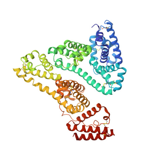
    Ruthenium(III) complexes are promising candidates for anticancer drugs, especially the clinically studied indazolium trans-[tetrachlorobis(1H-indazole)ruthenate(III)] (KP1019) and its analogue sodium trans-[tetrachlorobis(1H-indazole)ruthenate(III)] (NKP-1339). Several studies have emphasized the likely role of human serum proteins in the transportation and accumulation of ruthenium(III) complexes in tumors. Therefore, the interaction between KP1019 and human serum albumin was investigated by means of X-ray crystallography and inductively coupled plasma mass spectrometry (ICP-MS). The structural data unambiguously reveal the binding of two ruthenium atoms to histidine residues 146 and 242, which are both located within well-known hydrophobic binding pockets of albumin. The ruthenium centers are octahedrally coordinated by solvent molecules revealing the dissociation of both indazole ligands from the ruthenium-based drug. However, a binding mechanism is proposed indicating the importance of the indazole ligands for binding site recognition and thus their indispensable role for the binding of KP1019.


  • Organizational Affiliation
    • Fakultät für Chemie, Institut für Biophysikalische Chemie, Universität Wien , Althanstraße 14, 1090 Wien, Austria.

Macromolecule Content 

  • Total Structure Weight: 68.14 kDa 
  • Atom Count: 4,682 
  • Modeled Residue Count: 582 
  • Deposited Residue Count: 585 
  • Unique protein chains: 1

Macromolecules

Find similar proteins by:|  3D Structure
Entity ID: 1
MoleculeChains  Sequence LengthOrganismDetailsImage
Serum albumin585Homo sapiensMutation(s): 0 
UniProt & NIH Common Fund Data Resources
Find proteins for P02768 (Homo sapiens)
Explore P02768 
Go to UniProtKB:  P02768
PHAROS:  P02768
GTEx:  ENSG00000163631 
Entity Groups
Sequence Clusters30% Identity50% Identity70% Identity90% Identity95% Identity100% Identity
UniProt GroupP02768
Sequence Annotations
Expand
Reference Sequence

Small Molecules

Ligands 2 Unique
IDChains Name / Formula / InChI Key2D Diagram3D Interactions
MYR

Query on MYR


Download:Ideal Coordinates CCD File
D [auth A]
E [auth A]
F [auth A]
G [auth A]
H [auth A]
D [auth A],
E [auth A],
F [auth A],
G [auth A],
H [auth A],
I [auth A]
MYRISTIC ACID
C14 H28 O2
TUNFSRHWOTWDNC-UHFFFAOYSA-N
RU

Query on RU


Download:Ideal Coordinates CCD File
B [auth A],
C [auth A]
RUTHENIUM ION
Ru
BPEVHDGLPIIAGH-UHFFFAOYSA-N

Experimental Data & Validation

Experimental Data

  • Method: X-RAY DIFFRACTION
  • Resolution: 3.20 Å
  • R-Value Free:  0.262 (Depositor), 0.273 (DCC) 
  • R-Value Work:  0.244 (Depositor), 0.249 (DCC) 
  • R-Value Observed: 0.245 (Depositor) 
Space Group: C 1 2 1
Unit Cell:
Length ( Å )Angle ( ˚ )
a = 181.11α = 90
b = 38.06β = 105.06
c = 94.95γ = 90
Software Package:
Software NamePurpose
PHENIXrefinement
XDSdata scaling
PHENIXphasing
XDSdata reduction

Structure Validation

View Full Validation Report


Ligand Structure Quality Assessment 


Entry History 

& Funding Information

Deposition Data


Funding OrganizationLocationGrant Number
Austrian Science FundAustriaP23711

Revision History  (Full details and data files)

  • Version 1.0: 2016-06-01
    Type: Initial release
  • Version 1.1: 2016-07-06
    Changes: Database references
  • Version 1.2: 2017-09-06
    Changes: Author supporting evidence
  • Version 1.3: 2018-01-31
    Changes: Database references
  • Version 1.4: 2019-10-16
    Changes: Data collection
  • Version 1.5: 2024-01-10
    Changes: Data collection, Database references, Derived calculations, Refinement description
  • Version 1.6: 2024-11-06
    Changes: Structure summary