5IAA

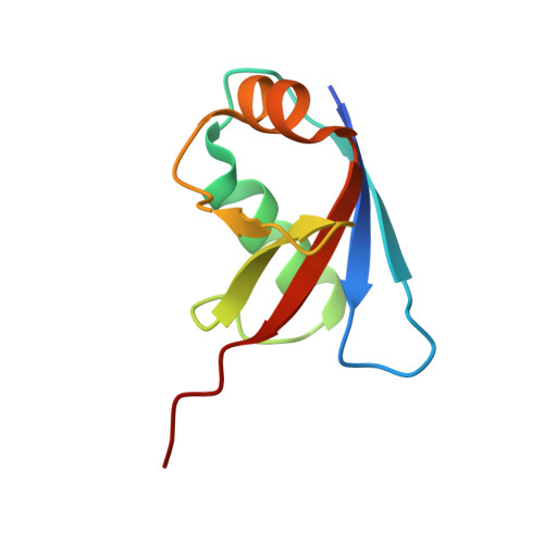
Crystal structure of human UBA5 in complex with UFM1


Experimental Data Snapshot

  • Method: X-RAY DIFFRACTION
  • Resolution: 1.85 Å
  • R-Value Free: 0.206 
  • R-Value Work: 0.187 
  • R-Value Observed: 0.188 

Starting Models: experimental
View more details

wwPDB Validation   3D Report Full Report


This is version 1.2 of the entry. See complete history


Literature

Trans-Binding Mechanism of Ubiquitin-like Protein Activation Revealed by a UBA5-UFM1 Complex.

Oweis, W.Padala, P.Hassouna, F.Cohen-Kfir, E.Gibbs, D.R.Todd, E.A.Berndsen, C.E.Wiener, R.

(2016) Cell Rep 16: 3113-3120

  • DOI: https://doi.org/10.1016/j.celrep.2016.08.067
  • Primary Citation of Related Structures:  
    5IAA, 5L95

  • PubMed Abstract: 

    Modification of proteins by ubiquitin or ubiquitin-like proteins (UBLs) is a critical cellular process implicated in a variety of cellular states and outcomes. A prerequisite for target protein modification by a UBL is the activation of the latter by activating enzymes (E1s). Here, we present the crystal structure of the non-canonical homodimeric E1, UBA5, in complex with its cognate UBL, UFM1, and supporting biochemical experiments. We find that UBA5 binds to UFM1 via a trans-binding mechanism in which UFM1 interacts with distinct sites in both subunits of the UBA5 dimer. This binding mechanism requires a region C-terminal to the adenylation domain that brings UFM1 to the active site of the adjacent UBA5 subunit. We also find that transfer of UFM1 from UBA5 to the E2, UFC1, occurs via a trans mechanism, thereby requiring a homodimer of UBA5. These findings explicitly elucidate the role of UBA5 dimerization in UFM1 activation.


  • Organizational Affiliation

    Department of Biochemistry and Molecular Biology, Institute for Medical Research Israel-Canada, Hebrew University-Hadassah Medical School, Jerusalem 91120, Israel.


Macromolecules
Find similar proteins by:  (by identity cutoff)  |  3D Structure
Entity ID: 1
MoleculeChains Sequence LengthOrganismDetailsImage
Ubiquitin-like modifier-activating enzyme 5
A, B
290Homo sapiensMutation(s): 0 
Gene Names: UBA5UBE1DC1
UniProt & NIH Common Fund Data Resources
Find proteins for Q9GZZ9 (Homo sapiens)
Explore Q9GZZ9 
Go to UniProtKB:  Q9GZZ9
PHAROS:  Q9GZZ9
GTEx:  ENSG00000081307 
Entity Groups  
Sequence Clusters30% Identity50% Identity70% Identity90% Identity95% Identity100% Identity
UniProt GroupQ9GZZ9
Sequence Annotations
Expand
  • Reference Sequence
Find similar proteins by:  (by identity cutoff)  |  3D Structure
Entity ID: 2
MoleculeChains Sequence LengthOrganismDetailsImage
Ubiquitin-fold modifier 1
C, D
83Homo sapiensMutation(s): 0 
Gene Names: UFM1C13orf20BM-002
UniProt & NIH Common Fund Data Resources
Find proteins for P61960 (Homo sapiens)
Explore P61960 
Go to UniProtKB:  P61960
PHAROS:  P61960
GTEx:  ENSG00000120686 
Entity Groups  
Sequence Clusters30% Identity50% Identity70% Identity90% Identity95% Identity100% Identity
UniProt GroupP61960
Sequence Annotations
Expand
  • Reference Sequence
Experimental Data & Validation

Experimental Data

  • Method: X-RAY DIFFRACTION
  • Resolution: 1.85 Å
  • R-Value Free: 0.206 
  • R-Value Work: 0.187 
  • R-Value Observed: 0.188 
  • Space Group: P 32 2 1
Unit Cell:
Length ( Å )Angle ( ˚ )
a = 138.081α = 90
b = 138.081β = 90
c = 99.178γ = 120
Software Package:
Software NamePurpose
REFMACrefinement
iMOSFLMdata reduction
SCALAdata scaling
PHASERphasing

Structure Validation

View Full Validation Report


Entry History & Funding Information

Deposition Data


Funding OrganizationLocationGrant Number
BSFIsrael--

Revision History  (Full details and data files)

  • Version 1.0: 2016-09-28
    Type: Initial release
  • Version 1.1: 2016-10-05
    Changes: Database references
  • Version 1.2: 2024-01-10
    Changes: Data collection, Database references, Refinement description