5I4C | pdb_00005i4c

Crystal structure of non-phosphorylated receiver domain of the stress response regulator RcsB from Escherichia coli


Experimental Data Snapshot

  • Method: X-RAY DIFFRACTION
  • Resolution: 2.00 Å
  • R-Value Free: 
    0.238 (Depositor), 0.230 (DCC) 
  • R-Value Work: 
    0.173 (Depositor), 0.181 (DCC) 
  • R-Value Observed: 
    0.176 (Depositor) 

wwPDB Validation 3D Report Full Report

Validation slider image for 5I4C

This is version 1.3 of the entry. See complete history

Literature

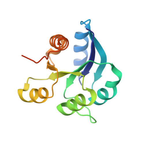
Crystal structure of nonphosphorylated receiver domain of the stress response regulator RcsB from Escherichia coli.

Filippova, E.V.Wawrzak, Z.Ruan, J.Pshenychnyi, S.Schultz, R.M.Wolfe, A.J.Anderson, W.F.

(2016) Protein Sci 25: 2216-2224

  • DOI: https://doi.org/10.1002/pro.3050
  • Primary Citation Related Structures: 
    5I4C

  • PubMed Abstract: 

    RcsB, the transcription-associated response regulator of the Rcs phosphorelay two-component signal transduction system, activates cell stress responses associated with desiccation, cell wall biosynthesis, cell division, virulence, biofilm formation, and antibiotic resistance in enteric bacterial pathogens. RcsB belongs to the FixJ/NarL family of transcriptional regulators, which are characterized by a highly conserved C-terminal DNA-binding domain. The N-terminal domain of RcsB belongs to the family of two-component receiver domains. This receiver domain contains the phosphoacceptor site and participates in RcsB dimer formation; it also contributes to dimer formation with other transcription factor partners. Here, we describe the crystal structure of the Escherichia coli RcsB receiver domain in its nonphosphorylated state. The structure reveals important molecular details of phosphorylation-independent dimerization of RcsB and has implication for the formation of heterodimers.


  • Organizational Affiliation
    • Department of Biochemistry and Molecular Genetics, Center for Structural Genomics of Infectious Diseases, Northwestern University Feinberg School of Medicine, Chicago, Illinois, 60611.

Macromolecule Content 

  • Total Structure Weight: 16.04 kDa 
  • Atom Count: 1,101 
  • Modeled Residue Count: 131 
  • Deposited Residue Count: 147 
  • Unique protein chains: 1

Macromolecules

Find similar proteins by:|  3D Structure
Entity ID: 1
MoleculeChains  Sequence LengthOrganismDetailsImage
Transcriptional regulatory protein RcsB147Escherichia coli K-12Mutation(s): 0 
Gene Names: rcsBb2217JW2205
UniProt
Find proteins for P0DMC7 (Escherichia coli (strain K12))
Explore P0DMC7 
Go to UniProtKB:  P0DMC7
Entity Groups
Sequence Clusters30% Identity50% Identity70% Identity90% Identity95% Identity100% Identity
UniProt GroupP0DMC7
Sequence Annotations
Expand
Reference Sequence

Experimental Data & Validation

Experimental Data

  • Method: X-RAY DIFFRACTION
  • Resolution: 2.00 Å
  • R-Value Free:  0.238 (Depositor), 0.230 (DCC) 
  • R-Value Work:  0.173 (Depositor), 0.181 (DCC) 
  • R-Value Observed: 0.176 (Depositor) 
Space Group: P 32 2 1
Unit Cell:
Length ( Å )Angle ( ˚ )
a = 61.197α = 90
b = 61.197β = 90
c = 65.226γ = 120
Software Package:
Software NamePurpose
REFMACrefinement
HKL-2000data scaling
PHENIXmodel building
BALBESphasing
HKL-2000data reduction

Structure Validation

View Full Validation Report


Entry History 

Revision History  (Full details and data files)

  • Version 1.0: 2016-10-12
    Type: Initial release
  • Version 1.1: 2016-11-30
    Changes: Database references
  • Version 1.2: 2017-11-01
    Changes: Author supporting evidence, Database references
  • Version 1.3: 2024-03-06
    Changes: Data collection, Database references