5I3B | pdb_00005i3b

Crystal Structure of tyrosinase from Bacillus megaterium with configuration B of hydroquinone inhibitor in the active site


Experimental Data Snapshot

  • Method: X-RAY DIFFRACTION
  • Resolution: 2.20 Å
  • R-Value Free: 
    0.217 (Depositor), 0.218 (DCC) 
  • R-Value Work: 
    0.186 (Depositor), 0.190 (DCC) 
  • R-Value Observed: 
    0.187 (Depositor) 

Starting Model: experimental
View more details

wwPDB Validation 3D Report Full Report

Validation slider image for 5I3B

This is version 1.2 of the entry. See complete history

Literature

The unravelling of the complex pattern of tyrosinase inhibition.

Deri, B.Kanteev, M.Goldfeder, M.Lecina, D.Guallar, V.Adir, N.Fishman, A.

(2016) Sci Rep 6: 34993-34993

  • DOI: https://doi.org/10.1038/srep34993
  • Primary Citation Related Structures: 
    5I38, 5I3A, 5I3B

  • PubMed Abstract: 

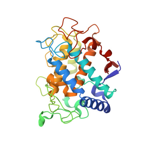
    Tyrosinases are responsible for melanin formation in all life domains. Tyrosinase inhibitors are used for the prevention of severe skin diseases, in skin-whitening creams and to avoid fruit browning, however continued use of many such inhibitors is considered unsafe. In this study we provide conclusive evidence of the inhibition mechanism of two well studied tyrosinase inhibitors, KA (kojic acid) and HQ (hydroquinone), which are extensively used in hyperpigmentation treatment. KA is reported in the literature with contradicting inhibition mechanisms, while HQ is described as both a tyrosinase inhibitor and a substrate. By visualization of KA and HQ in the active site of TyrBm crystals, together with molecular modeling, binding constant analysis and kinetic experiments, we have elucidated their mechanisms of inhibition, which was ambiguous for both inhibitors. We confirm that while KA acts as a mixed inhibitor, HQ can act both as a TyrBm substrate and as an inhibitor.


  • Organizational Affiliation
    • Department of Biotechnology and Food Engineering, Technion-Israel Institute of Technology, Haifa, 3200003, Israel.

Macromolecule Content 

  • Total Structure Weight: 67.13 kDa 
  • Atom Count: 4,995 
  • Modeled Residue Count: 574 
  • Deposited Residue Count: 574 
  • Unique protein chains: 1

Macromolecules

Find similar proteins by:|  3D Structure
Entity ID: 1
MoleculeChains  Sequence LengthOrganismDetailsImage
Tyrosinase
A, B
287Priestia megateriumMutation(s): 0 
UniProt
Find proteins for B2ZB02 (Priestia megaterium)
Explore B2ZB02 
Go to UniProtKB:  B2ZB02
Entity Groups
Sequence Clusters30% Identity50% Identity70% Identity90% Identity95% Identity100% Identity
UniProt GroupB2ZB02
Sequence Annotations
Expand
Reference Sequence

Experimental Data & Validation

Experimental Data

  • Method: X-RAY DIFFRACTION
  • Resolution: 2.20 Å
  • R-Value Free:  0.217 (Depositor), 0.218 (DCC) 
  • R-Value Work:  0.186 (Depositor), 0.190 (DCC) 
  • R-Value Observed: 0.187 (Depositor) 
Space Group: P 21 21 21
Unit Cell:
Length ( Å )Angle ( ˚ )
a = 69.62α = 90
b = 74.42β = 90
c = 119.69γ = 90
Software Package:
Software NamePurpose
PHENIXrefinement
MOSFLMdata reduction
SCALAdata scaling
PHASERphasing
PDB_EXTRACTdata extraction

Structure Validation

View Full Validation Report


Entry History 

& Funding Information

Deposition Data


Funding OrganizationLocationGrant Number
ISFIsrael419/15

Revision History  (Full details and data files)

  • Version 1.0: 2016-10-12
    Type: Initial release
  • Version 1.1: 2016-10-19
    Changes: Database references
  • Version 1.2: 2024-01-10
    Changes: Data collection, Database references, Derived calculations, Refinement description