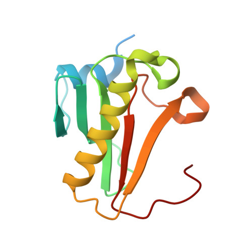
5I2F

Human Histidine Triad Nucleotide Binding Protein 1 (hHint1) with bound sulfamide inhibitor Bio-AMS


Experimental Data Snapshot

  • Method: X-RAY DIFFRACTION
  • Resolution: 1.25 Å
  • R-Value Free: 0.175 
  • R-Value Work: 0.155 
  • R-Value Observed: 0.156 

Starting Model: experimental
View more details

wwPDB Validation   3D Report Full Report


Ligand Structure Quality Assessment 


This is version 1.2 of the entry. See complete history


Literature

Design, Synthesis, and Characterization of Sulfamide and Sulfamate Nucleotidomimetic Inhibitors of hHint1.

Shah, R.Strom, A.Zhou, A.Maize, K.M.Finzel, B.C.Wagner, C.R.

(2016) ACS Med Chem Lett 7: 780-784

  • DOI: https://doi.org/10.1021/acsmedchemlett.6b00169
  • Primary Citation of Related Structures:  
    5I2E, 5I2F

  • PubMed Abstract: 

    Hint1 has recently emerged to be an important target of interest due to its involvement in the regulation of a broad range of CNS functions including opioid signaling, tolerance, neuropathic pain, and nicotine dependence. A series of inhibitors were rationally designed, synthesized, and tested for their inhibitory activity against hHint1 using isothermal titration calorimetry (ITC). The studies resulted in the development of the first small-molecule inhibitors of hHint1 with submicromolar binding affinities. A combination of thermodynamic and high-resolution X-ray crystallographic studies provides an insight into the biomolecular recognition of ligands by hHint1. These novel inhibitors have potential utility as molecular probes to better understand the role and function of hHint1 in the CNS.


  • Organizational Affiliation

    Departments of Medicinal Chemistry and Chemistry, University of Minnesota , Minneapolis, Minnesota 55455, United States.


Macromolecules
Find similar proteins by:  (by identity cutoff)  |  3D Structure
Entity ID: 1
MoleculeChains Sequence LengthOrganismDetailsImage
Histidine triad nucleotide-binding protein 1A [auth B],
B [auth A]
129Homo sapiensMutation(s): 0 
Gene Names: HINT1HINT
EC: 3 (PDB Primary Data), 3.4.22 (UniProt), 3.9.1 (UniProt)
UniProt & NIH Common Fund Data Resources
Find proteins for P49773 (Homo sapiens)
Explore P49773 
Go to UniProtKB:  P49773
PHAROS:  P49773
GTEx:  ENSG00000169567 
Entity Groups  
Sequence Clusters30% Identity50% Identity70% Identity90% Identity95% Identity100% Identity
UniProt GroupP49773
Sequence Annotations
Expand
  • Reference Sequence
Small Molecules
Binding Affinity Annotations 
IDSourceBinding Affinity
BS5 BindingDB:  5I2F Kd: 320 (nM) from 1 assay(s)
IC50: 1000 (nM) from 1 assay(s)
Experimental Data & Validation

Experimental Data

  • Method: X-RAY DIFFRACTION
  • Resolution: 1.25 Å
  • R-Value Free: 0.175 
  • R-Value Work: 0.155 
  • R-Value Observed: 0.156 
  • Space Group: C 1 2 1
Unit Cell:
Length ( Å )Angle ( ˚ )
a = 77.493α = 90
b = 46.314β = 94.44
c = 64.073γ = 90
Software Package:
Software NamePurpose
REFMACrefinement
PDB_EXTRACTdata extraction
PHASERphasing
SCALAdata scaling
XDSdata reduction

Structure Validation

View Full Validation Report


Ligand Structure Quality Assessment 


Entry History 

Deposition Data

Revision History  (Full details and data files)

  • Version 1.0: 2016-06-22
    Type: Initial release
  • Version 1.1: 2016-09-14
    Changes: Database references
  • Version 1.2: 2023-09-27
    Changes: Data collection, Database references, Derived calculations, Refinement description