5I1W | pdb_00005i1w

Crystal structure of CrmK, a flavoenzyme involved in the shunt product recycling mechanism in caerulomycin biosynthesis


Experimental Data Snapshot

  • Method: X-RAY DIFFRACTION
  • Resolution: 2.15 Å
  • R-Value Free: 
    0.213 (Depositor), 0.218 (DCC) 
  • R-Value Work: 
    0.168 (Depositor), 0.176 (DCC) 
  • R-Value Observed: 
    0.171 (Depositor) 

Starting Model: experimental
View more details

wwPDB Validation 3D Report Full Report

Validation slider image for 5I1W

Ligand Structure Quality Assessment 


This is version 1.4 of the entry. See complete history

Literature

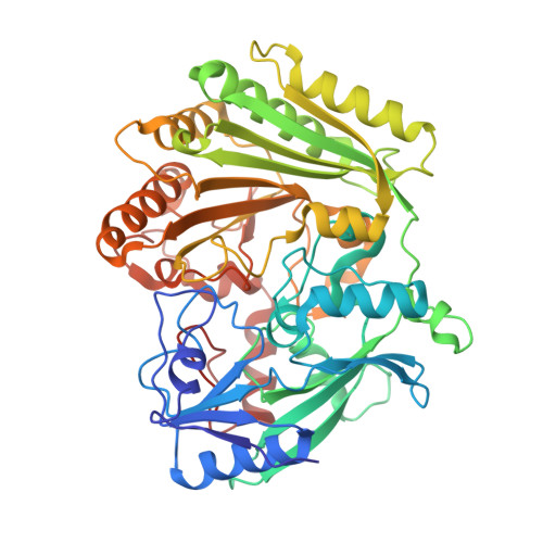
Biochemical and structural insights into flavoenzyme CrmK reveals a shunt product recycling mechanism in caerulomycin biosynthesis

Zhu, Y.Picard, M.-E.Mei, X.Zhang, Q.Barma, J.Murphy Despres, X.Couture, M.Zhu, W.Zhang, C.Shi, R.

To be published.

Macromolecule Content 

  • Total Structure Weight: 225.09 kDa 
  • Atom Count: 16,449 
  • Modeled Residue Count: 1,992 
  • Deposited Residue Count: 2,000 
  • Unique protein chains: 1

Macromolecules

Find similar proteins by:|  3D Structure
Entity ID: 1
MoleculeChains  Sequence LengthOrganismDetailsImage
CrmK
A, B, C, D
500Actinoalloteichus sp. WH1-2216-6Mutation(s): 0 
UniProt
Find proteins for H8Y6P5 (Actinoalloteichus sp. WH1-2216-6)
Explore H8Y6P5 
Go to UniProtKB:  H8Y6P5
Entity Groups
Sequence Clusters30% Identity50% Identity70% Identity90% Identity95% Identity100% Identity
UniProt GroupH8Y6P5
Sequence Annotations
Expand
Reference Sequence

Small Molecules

Ligands 3 Unique
IDChains Name / Formula / InChI Key2D Diagram3D Interactions
FAD

Query on FAD


Download:Ideal Coordinates CCD File
E [auth A],
G [auth B],
I [auth C],
K [auth D]
FLAVIN-ADENINE DINUCLEOTIDE
C27 H33 N9 O15 P2
VWWQXMAJTJZDQX-UYBVJOGSSA-N
67L

Query on 67L


Download:Ideal Coordinates CCD File
F [auth A],
H [auth B],
J [auth C]
6-(hydroxymethyl)[2,2'-bipyridin]-4-ol
C11 H10 N2 O2
HKHZGZONLACWBE-UHFFFAOYSA-N
67K

Query on 67K


Download:Ideal Coordinates CCD File
L [auth D]4-hydroxy[2,2'-bipyridine]-6-carbaldehyde
C11 H8 N2 O2
FFOWHRKNAPHKGV-UHFFFAOYSA-N

Experimental Data & Validation

Experimental Data

  • Method: X-RAY DIFFRACTION
  • Resolution: 2.15 Å
  • R-Value Free:  0.213 (Depositor), 0.218 (DCC) 
  • R-Value Work:  0.168 (Depositor), 0.176 (DCC) 
  • R-Value Observed: 0.171 (Depositor) 
Space Group: P 1
Unit Cell:
Length ( Å )Angle ( ˚ )
a = 63.11α = 94.73
b = 95.2β = 96.56
c = 98.04γ = 105.2
Software Package:
Software NamePurpose
MOSFLMdata reduction
SCALAdata scaling
REFMACrefinement
PDB_EXTRACTdata extraction

Structure Validation

View Full Validation Report


Ligand Structure Quality Assessment 


Entry History 

& Funding Information

Deposition Data


Funding OrganizationLocationGrant Number
Natural Sciences and Engineering Research Council (NSERC, Canada)Canada--

Revision History  (Full details and data files)

  • Version 1.0: 2017-02-15
    Type: Initial release
  • Version 1.1: 2017-09-13
    Changes: Author supporting evidence
  • Version 1.2: 2020-01-08
    Changes: Author supporting evidence, Derived calculations
  • Version 1.3: 2023-09-27
    Changes: Data collection, Database references, Derived calculations, Refinement description
  • Version 1.4: 2024-10-23
    Changes: Structure summary