5HSA | pdb_00005hsa

Alcohol Oxidase AOX1 from Pichia Pastoris


Experimental Data Snapshot

  • Method: X-RAY DIFFRACTION
  • Resolution: 2.35 Å
  • R-Value Free: 
    0.205 (Depositor), 0.207 (DCC) 
  • R-Value Work: 
    0.177 (Depositor), 0.182 (DCC) 
  • R-Value Observed: 
    0.179 (Depositor) 

Starting Model: experimental
View more details

wwPDB Validation 3D Report Full Report

Validation slider image for 5HSA

Ligand Structure Quality Assessment 


This is version 1.1 of the entry. See complete history

Literature

Crystal Structure of Alcohol Oxidase from Pichia pastoris.

Koch, C.Neumann, P.Valerius, O.Feussner, I.Ficner, R.

(2016) PLoS One 11: e0149846-e0149846

  • DOI: https://doi.org/10.1371/journal.pone.0149846
  • Primary Citation Related Structures: 
    5HSA

  • PubMed Abstract: 

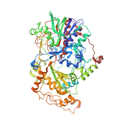
    FAD-dependent alcohol oxidases (AOX) are key enzymes of methylotrophic organisms that can utilize lower primary alcohols as sole source of carbon and energy. Here we report the crystal structure analysis of the methanol oxidase AOX1 from Pichia pastoris. The crystallographic phase problem was solved by means of Molecular Replacement in combination with initial structure rebuilding using Rosetta model completion and relaxation against an averaged electron density map. The subunit arrangement of the homo-octameric AOX1 differs from that of octameric vanillyl alcohol oxidase and other dimeric or tetrameric alcohol oxidases, due to the insertion of two large protruding loop regions and an additional C-terminal extension in AOX1. In comparison to other alcohol oxidases, the active site cavity of AOX1 is significantly reduced in size, which could explain the observed preference for methanol as substrate. All AOX1 subunits of the structure reported here harbor a modified flavin adenine dinucleotide, which contains an arabityl chain instead of a ribityl chain attached to the isoalloxazine ring.


  • Organizational Affiliation
    • Department of Plant Biochemistry, Albrecht-von-Haller-Institute, Georg-August-University Goettingen, Justus-von-Liebig-Weg 11, 37077, Goettingen, Germany.

Macromolecule Content 

  • Total Structure Weight: 602.84 kDa 
  • Atom Count: 44,407 
  • Modeled Residue Count: 5,296 
  • Deposited Residue Count: 5,304 
  • Unique protein chains: 1

Macromolecules

Find similar proteins by:|  3D Structure
Entity ID: 1
MoleculeChains  Sequence LengthOrganismDetailsImage
Alcohol oxidase 1
A, B, C, D, E
A, B, C, D, E, F, G, H
663Komagataella phaffii CBS 7435Mutation(s): 0 
Gene Names: AOX1PP7435_Chr4-0130
EC: 1.1.3.13
UniProt
Find proteins for F2QY27 (Komagataella phaffii (strain ATCC 76273 / CBS 7435 / CECT 11047 / NRRL Y-11430 / Wegner 21-1))
Explore F2QY27 
Go to UniProtKB:  F2QY27
Entity Groups
Sequence Clusters30% Identity50% Identity70% Identity90% Identity95% Identity100% Identity
UniProt GroupF2QY27
Sequence Annotations
Expand
Reference Sequence

Small Molecules

Ligands 7 Unique
IDChains Name / Formula / InChI Key2D Diagram3D Interactions
FAS

Query on FAS


Download:Ideal Coordinates CCD File
BA [auth D]
FA [auth E]
I [auth A]
JA [auth F]
N [auth B]
BA [auth D],
FA [auth E],
I [auth A],
JA [auth F],
N [auth B],
PA [auth G],
T [auth C],
TA [auth H]
ARABINO-FLAVIN-ADENINE DINUCLEOTIDE
C27 H33 N9 O15 P2
VWWQXMAJTJZDQX-WCVIGVMNSA-N
P6G

Query on P6G


Download:Ideal Coordinates CCD File
CA [auth D]
GA [auth E]
J [auth A]
KA [auth F]
LA [auth F]
CA [auth D],
GA [auth E],
J [auth A],
KA [auth F],
LA [auth F],
O [auth B],
P [auth B],
U [auth C],
V [auth C]
HEXAETHYLENE GLYCOL
C12 H26 O7
IIRDTKBZINWQAW-UHFFFAOYSA-N
PGE

Query on PGE


Download:Ideal Coordinates CCD File
DA [auth D]
HA [auth E]
K [auth A]
Q [auth B]
QA [auth G]
DA [auth D],
HA [auth E],
K [auth A],
Q [auth B],
QA [auth G],
R [auth B],
RA [auth G],
UA [auth H],
W [auth C]
TRIETHYLENE GLYCOL
C6 H14 O4
ZIBGPFATKBEMQZ-UHFFFAOYSA-N
PO4

Query on PO4


Download:Ideal Coordinates CCD File
L [auth A],
MA [auth F]
PHOSPHATE ION
O4 P
NBIIXXVUZAFLBC-UHFFFAOYSA-K
GOL

Query on GOL


Download:Ideal Coordinates CCD File
NA [auth F],
X [auth C]
GLYCEROL
C3 H8 O3
PEDCQBHIVMGVHV-UHFFFAOYSA-N
CA

Query on CA


Download:Ideal Coordinates CCD File
AA [auth C]
EA [auth D]
IA [auth E]
M [auth A]
OA [auth F]
AA [auth C],
EA [auth D],
IA [auth E],
M [auth A],
OA [auth F],
S [auth B],
SA [auth G],
Z [auth C]
CALCIUM ION
Ca
BHPQYMZQTOCNFJ-UHFFFAOYSA-N
CL

Query on CL


Download:Ideal Coordinates CCD File
Y [auth C]CHLORIDE ION
Cl
VEXZGXHMUGYJMC-UHFFFAOYSA-M

Experimental Data & Validation

Experimental Data

  • Method: X-RAY DIFFRACTION
  • Resolution: 2.35 Å
  • R-Value Free:  0.205 (Depositor), 0.207 (DCC) 
  • R-Value Work:  0.177 (Depositor), 0.182 (DCC) 
  • R-Value Observed: 0.179 (Depositor) 
Space Group: P 1 21 1
Unit Cell:
Length ( Å )Angle ( ˚ )
a = 117.1α = 90
b = 165.19β = 95.67
c = 164.31γ = 90
Software Package:
Software NamePurpose
XSCALEdata scaling
PHASERphasing
PHENIXrefinement
PDB_EXTRACTdata extraction
XDSdata reduction

Structure Validation

View Full Validation Report


Ligand Structure Quality Assessment 


Entry History 

& Funding Information

Deposition Data


Funding OrganizationLocationGrant Number
German Research FoundationGermanyIRTG 1422
Goettingen UniversityGermanyOpen Access Publication Funds

Revision History  (Full details and data files)

  • Version 1.0: 2016-03-09
    Type: Initial release
  • Version 1.1: 2024-01-10
    Changes: Author supporting evidence, Data collection, Database references, Derived calculations, Refinement description