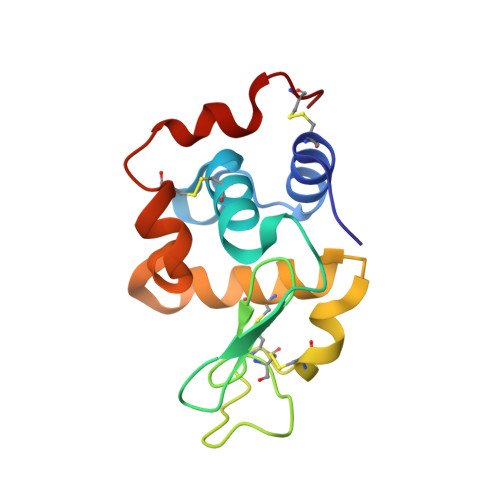
5HNL

In-house X-ray single crystal diffraction from protein microcrystals via magnetically oriented microcrystal arrays in gels


Experimental Data Snapshot

  • Method: X-RAY DIFFRACTION
  • Resolution: 2.42 Å
  • R-Value Free: 0.188 
  • R-Value Work: 0.171 
  • R-Value Observed: 0.172 

wwPDB Validation   3D Report Full Report


This is version 1.1 of the entry. See complete history


Literature

Neutron and X-ray single-crystal diffraction from protein microcrystals via magnetically oriented microcrystal arrays in gels.

Tsukui, S.Kimura, F.Kusaka, K.Baba, S.Mizuno, N.Kimura, T.

(2016) Acta Crystallogr D Struct Biol 72: 823-829

  • DOI: https://doi.org/10.1107/S2059798316007415
  • Primary Citation of Related Structures:  
    5HNC, 5HNL

  • PubMed Abstract: 

    Protein microcrystals magnetically aligned in D2O hydrogels were subjected to neutron diffraction measurements, and reflections were observed for the first time to a resolution of 3.4 Å from lysozyme microcrystals (∼10 × 10 × 50 µm). This result demonstrated the possibility that magnetically oriented microcrystals consolidated in D2O gels may provide a promising means to obtain single-crystal neutron diffraction from proteins that do not crystallize at the sizes required for neutron diffraction structure determination. In addition, lysozyme microcrystals aligned in H2O hydrogels allowed structure determination at a resolution of 1.76 Å at room temperature by X-ray diffraction. The use of gels has advantages since the microcrystals are measured under hydrated conditions.


  • Organizational Affiliation

    Division of Forest and Biomaterials Science, Graduate School of Agriculture, Kyoto University, Kitashirakawa, Sakyo-ku, Kyoto 606-8502, Japan.


Macromolecules
Find similar proteins by:  (by identity cutoff)  |  3D Structure
Entity ID: 1
MoleculeChains Sequence LengthOrganismDetailsImage
Lysozyme C129Gallus gallusMutation(s): 0 
EC: 3.2.1.17
UniProt
Find proteins for P00698 (Gallus gallus)
Explore P00698 
Go to UniProtKB:  P00698
Entity Groups  
Sequence Clusters30% Identity50% Identity70% Identity90% Identity95% Identity100% Identity
UniProt GroupP00698
Sequence Annotations
Expand
  • Reference Sequence
Experimental Data & Validation

Experimental Data

  • Method: X-RAY DIFFRACTION
  • Resolution: 2.42 Å
  • R-Value Free: 0.188 
  • R-Value Work: 0.171 
  • R-Value Observed: 0.172 
  • Space Group: P 21 21 21
Unit Cell:
Length ( Å )Angle ( ˚ )
a = 30.53α = 90
b = 56.42β = 90
c = 73.34γ = 90
Software Package:
Software NamePurpose
PHENIXrefinement
iMOSFLMdata reduction
Aimlessdata scaling
PHASERphasing

Structure Validation

View Full Validation Report


Entry History 

Deposition Data

Revision History  (Full details and data files)

  • Version 1.0: 2016-07-20
    Type: Initial release
  • Version 1.1: 2024-10-30
    Changes: Data collection, Database references, Derived calculations, Structure summary