5HLY | pdb_00005hly

Structure of Pro-Activin A Precursor at 2.3 A Resolution


Experimental Data Snapshot

  • Method: X-RAY DIFFRACTION
  • Resolution: 2.30 Å
  • R-Value Free: 
    0.252 (Depositor), 0.255 (DCC) 
  • R-Value Work: 
    0.203 (Depositor), 0.204 (DCC) 
  • R-Value Observed: 
    0.205 (Depositor) 

Starting Model: experimental
View more details

wwPDB Validation 3D Report Full Report

Validation slider image for 5HLY

This is version 1.2 of the entry. See complete history

Literature

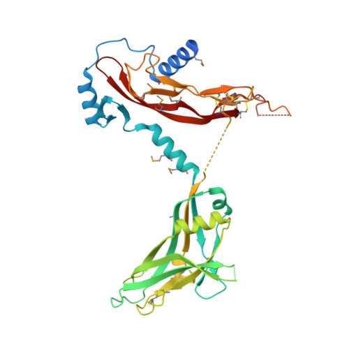
Structure and activation of pro-activin A.

Wang, X.Fischer, G.Hyvonen, M.

(2016) Nat Commun 7: 12052-12052

  • DOI: https://doi.org/10.1038/ncomms12052
  • Primary Citation Related Structures: 
    5HLY, 5HLZ

  • PubMed Abstract: 

    Activins are growth factors with multiple roles in the development and homeostasis. Like all TGF-β family of growth factors, activins are synthesized as large precursors from which mature dimeric growth factors are released proteolytically. Here we have studied the activation of activin A and determined crystal structures of the unprocessed precursor and of the cleaved pro-mature complex. Replacing the natural furin cleavage site with a HRV 3C protease site, we show how the protein gains its bioactivity after proteolysis and is as active as the isolated mature domain. The complex remains associated in conditions used for biochemical analysis with a dissociation constant of 5 nM, but the pro-domain can be actively displaced from the complex by follistatin. Our high-resolution structures of pro-activin A share features seen in the pro-TGF-β1 and pro-BMP-9 structures, but reveal a new oligomeric arrangement, with a domain-swapped, cross-armed conformation for the protomers in the dimeric protein.


  • Organizational Affiliation
    • Department of Biochemistry, University of Cambridge, 80 Tennis Court Road, Cambridge CB2 1GA, UK.

Macromolecule Content 

  • Total Structure Weight: 43.7 kDa 
  • Atom Count: 2,406 
  • Modeled Residue Count: 299 
  • Deposited Residue Count: 383 
  • Unique protein chains: 1

Macromolecules

Find similar proteins by:|  3D Structure
Entity ID: 1
MoleculeChains  Sequence LengthOrganismDetailsImage
Inhibin beta A chain383Homo sapiensMutation(s): 2 
Gene Names: INHBA
UniProt & NIH Common Fund Data Resources
Find proteins for P08476 (Homo sapiens)
Explore P08476 
Go to UniProtKB:  P08476
PHAROS:  P08476
GTEx:  ENSG00000122641 
Entity Groups
Sequence Clusters30% Identity50% Identity70% Identity90% Identity95% Identity100% Identity
UniProt GroupP08476
Sequence Annotations
Expand
Reference Sequence

Small Molecules

Ligands 1 Unique
IDChains Name / Formula / InChI Key2D Diagram3D Interactions
CL

Query on CL


Download:Ideal Coordinates CCD File
B [auth A]CHLORIDE ION
Cl
VEXZGXHMUGYJMC-UHFFFAOYSA-M
Modified Residues  1 Unique
IDChains TypeFormula2D DiagramParent
MSE
Query on MSE
A
L-PEPTIDE LINKINGC5 H11 N O2 SeMET

Experimental Data & Validation

Experimental Data

  • Method: X-RAY DIFFRACTION
  • Resolution: 2.30 Å
  • R-Value Free:  0.252 (Depositor), 0.255 (DCC) 
  • R-Value Work:  0.203 (Depositor), 0.204 (DCC) 
  • R-Value Observed: 0.205 (Depositor) 
Space Group: P 61 2 2
Unit Cell:
Length ( Å )Angle ( ˚ )
a = 46.561α = 90
b = 46.561β = 90
c = 444.921γ = 120
Software Package:
Software NamePurpose
PHENIXrefinement
XDSdata reduction
Aimlessdata scaling
PHASERphasing
Cootmodel building

Structure Validation

View Full Validation Report


Entry History 

Deposition Data

Revision History  (Full details and data files)

  • Version 1.0: 2016-07-13
    Type: Initial release
  • Version 1.1: 2024-05-01
    Changes: Data collection, Database references, Refinement description
  • Version 1.2: 2024-11-20
    Changes: Structure summary