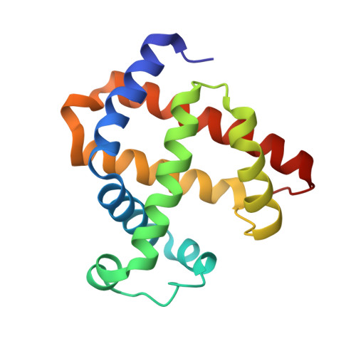
Regulating the nitrite reductase activity of myoglobin by redesigning the heme active center
Wu, L.B., Yuan, H., Gao, S.Q., You, Y., Nie, C.M., Wen, G.B., Lin, Y.W., Tan, X.(2016) Nitric Oxide 57: 21-29
- PubMed: 27108710
- DOI: https://doi.org/10.1016/j.niox.2016.04.007
- Primary Citation of Related Structures:
5HLQ, 5HLU, 5HLX - PubMed Abstract:
Heme proteins perform diverse functions in living systems, of which nitrite reductase (NIR) activity receives much attention recently. In this study, to better understand the structural elements responsible for the NIR activity, we used myoglobin (Mb) as a model heme protein and redesigned the heme active center, by introducing one or two distal histidines, and by creating a channel to the heme center with removal of the native distal His64 gate (His to Ala mutation). UV-Vis kinetic studies, combined with EPR studies, showed that a single distal histidine with a suitable position to the heme iron, i.e., His43, is crucial for nitrite (NO2(-)) to nitric oxide (NO) reduction. Moreover, creation of a water channel to the heme center significantly enhanced the NIR activity compared to the corresponding mutant without the channel. In addition, X-ray crystallographic studies of F43H/H64A Mb and its complexes with NO2(-) or NO revealed a unique hydrogen-bonding network in the heme active center, as well as unique substrate and product binding models, providing valuable structural information for the enhanced NIR activity. These findings enriched our understanding of the structure and NIR activity relationship of heme proteins. The approach of creating a channel in this study is also useful for rational design of other functional heme proteins.
- School of Chemistry and Chemical Engineering, University of South China, Hengyang 421001, China.
Organizational Affiliation:


