5HIU | pdb_00005hiu

Structure of the TSC2 N-terminus


Experimental Data Snapshot

  • Method: X-RAY DIFFRACTION
  • Resolution: 2.50 Å
  • R-Value Free: 
    0.263 (Depositor), 0.263 (DCC) 
  • R-Value Work: 
    0.224 (Depositor), 0.233 (DCC) 
  • R-Value Observed: 
    0.226 (Depositor) 

wwPDB Validation 3D Report Full Report

Validation slider image for 5HIU

This is version 1.3 of the entry. See complete history

Literature

Structure of the Tuberous Sclerosis Complex 2 (TSC2) N Terminus Provides Insight into Complex Assembly and Tuberous Sclerosis Pathogenesis.

Zech, R.Kiontke, S.Mueller, U.Oeckinghaus, A.Kummel, D.

(2016) J Biological Chem 291: 20008-20020

  • DOI: https://doi.org/10.1074/jbc.M116.732446
  • Primary Citation Related Structures: 
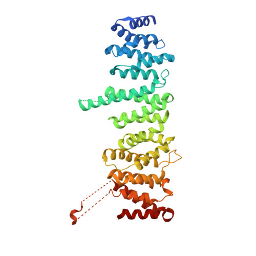
    5HIU

  • PubMed Abstract: 

    Tuberous sclerosis complex (TSC) is caused by mutations in the TSC1 and TSC2 tumor suppressor genes. The gene products hamartin and tuberin form the TSC complex that acts as GTPase-activating protein for Rheb and negatively regulates the mammalian target of rapamycin complex 1 (mTORC1). Tuberin contains a RapGAP homology domain responsible for inactivation of Rheb, but functions of other protein domains remain elusive. Here we show that the TSC2 N terminus interacts with the TSC1 C terminus to mediate complex formation. The structure of the TSC2 N-terminal domain from Chaetomium thermophilum and a homology model of the human tuberin N terminus are presented. We characterize the molecular requirements for TSC1-TSC2 interactions and analyze pathological point mutations in tuberin. Many mutations are structural and produce improperly folded protein, explaining their effect in pathology, but we identify one point mutant that abrogates complex formation without affecting protein structure. We provide the first structural information on TSC2/tuberin with novel insight into the molecular function.


  • Organizational Affiliation
    • From the Structural Biology Section, FB5 Biology/Chemistry, University of Osnabrück, 49076 Osnabrück, Germany.

Macromolecule Content 

  • Total Structure Weight: 206.19 kDa 
  • Atom Count: 12,812 
  • Modeled Residue Count: 1,617 
  • Deposited Residue Count: 1,852 
  • Unique protein chains: 1

Macromolecules

Find similar proteins by:|  3D Structure
Entity ID: 1
MoleculeChains  Sequence LengthOrganismDetailsImage
GTPase activator-like protein
A, B, C, D
463Thermochaetoides thermophilaMutation(s): 0 
Gene Names: CTHT_0061860
UniProt
Find proteins for G0SFF5 (Chaetomium thermophilum (strain DSM 1495 / CBS 144.50 / IMI 039719))
Explore G0SFF5 
Go to UniProtKB:  G0SFF5
Entity Groups
Sequence Clusters30% Identity50% Identity70% Identity90% Identity95% Identity100% Identity
UniProt GroupG0SFF5
Sequence Annotations
Expand
Reference Sequence

Experimental Data & Validation

Experimental Data

  • Method: X-RAY DIFFRACTION
  • Resolution: 2.50 Å
  • R-Value Free:  0.263 (Depositor), 0.263 (DCC) 
  • R-Value Work:  0.224 (Depositor), 0.233 (DCC) 
  • R-Value Observed: 0.226 (Depositor) 
Space Group: P 1
Unit Cell:
Length ( Å )Angle ( ˚ )
a = 42.73α = 89.9
b = 78.1β = 89.85
c = 153.75γ = 78.94
Software Package:
Software NamePurpose
REFMACrefinement
XDSdata reduction
SCALAdata scaling
AutoSolphasing

Structure Validation

View Full Validation Report


Entry History 

& Funding Information

Deposition Data


Funding OrganizationLocationGrant Number
German Research FoundationGermanyKU 2531/2-1

Revision History  (Full details and data files)

  • Version 1.0: 2016-08-10
    Type: Initial release
  • Version 1.1: 2016-08-17
    Changes: Database references
  • Version 1.2: 2016-09-28
    Changes: Database references
  • Version 1.3: 2024-05-08
    Changes: Author supporting evidence, Data collection, Database references, Refinement description