5HH3 | pdb_00005hh3

OxyA from Actinoplanes teichomyceticus


Experimental Data Snapshot

  • Method: X-RAY DIFFRACTION
  • Resolution: 2.10 Å
  • R-Value Free: 
    0.229 (Depositor), 0.235 (DCC) 
  • R-Value Work: 
    0.198 (Depositor), 0.205 (DCC) 
  • R-Value Observed: 
    0.200 (Depositor) 

Starting Model: experimental
View more details

wwPDB Validation 3D Report Full Report

Validation slider image for 5HH3

Ligand Structure Quality Assessment 


This is version 1.3 of the entry. See complete history

Literature

Structure of OxyAtei : completing our picture of the glycopeptide antibiotic producing Cytochrome P450 cascade.

Haslinger, K.Cryle, M.J.

(2016) FEBS Lett 590: 571-581

  • DOI: https://doi.org/10.1002/1873-3468.12081
  • Primary Citation Related Structures: 
    5HH3

  • PubMed Abstract: 

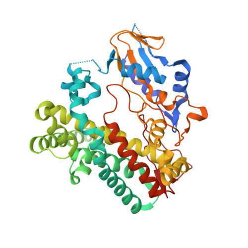
    Cyclization of glycopeptide antibiotic precursors occurs in either three or four steps catalyzed by Cytochrome P450 enzymes. Three of these enzymes have been structurally characterized to date with the second enzyme along the pathway, OxyA, escaping structural analysis. We are now able to present the structure of OxyAtei involved in teicoplanin biosynthesis - the same enzyme recently shown to be the first active OxyA homolog. In spite of the hydrophobic character of the teicoplanin precursor, the polar active site of OxyAtei and its affinity for certain azole inhibitors hint at its preference for substrates with polar decorations.


  • Organizational Affiliation
    • Max Planck Institute for Medical Research, Heidelberg, Germany.

Macromolecule Content 

  • Total Structure Weight: 91.19 kDa 
  • Atom Count: 6,471 
  • Modeled Residue Count: 774 
  • Deposited Residue Count: 794 
  • Unique protein chains: 1

Macromolecules

Find similar proteins by:|  3D Structure
Entity ID: 1
MoleculeChains  Sequence LengthOrganismDetailsImage
OxyA proteinA,
B [auth C]
397Actinoplanes teichomyceticusMutation(s): 0 
Gene Names: tcp18
UniProt
Find proteins for Q6ZZI8 (Actinoplanes teichomyceticus)
Explore Q6ZZI8 
Go to UniProtKB:  Q6ZZI8
Entity Groups
Sequence Clusters30% Identity50% Identity70% Identity90% Identity95% Identity100% Identity
UniProt GroupQ6ZZI8
Sequence Annotations
Expand
Reference Sequence

Small Molecules

Ligands 3 Unique
IDChains Name / Formula / InChI Key2D Diagram3D Interactions
HEM

Query on HEM


Download:Ideal Coordinates CCD File
C [auth A],
G [auth C]
PROTOPORPHYRIN IX CONTAINING FE
C34 H32 Fe N4 O4
KABFMIBPWCXCRK-RGGAHWMASA-L
GOL

Query on GOL


Download:Ideal Coordinates CCD File
D [auth A]
E [auth A]
H [auth C]
I [auth C]
J [auth C]
D [auth A],
E [auth A],
H [auth C],
I [auth C],
J [auth C],
K [auth C],
L [auth C],
M [auth C],
N [auth C]
GLYCEROL
C3 H8 O3
PEDCQBHIVMGVHV-UHFFFAOYSA-N
ACT

Query on ACT


Download:Ideal Coordinates CCD File
F [auth A],
O [auth C],
P [auth C]
ACETATE ION
C2 H3 O2
QTBSBXVTEAMEQO-UHFFFAOYSA-M

Experimental Data & Validation

Experimental Data

  • Method: X-RAY DIFFRACTION
  • Resolution: 2.10 Å
  • R-Value Free:  0.229 (Depositor), 0.235 (DCC) 
  • R-Value Work:  0.198 (Depositor), 0.205 (DCC) 
  • R-Value Observed: 0.200 (Depositor) 
Space Group: P 21 21 21
Unit Cell:
Length ( Å )Angle ( ˚ )
a = 58.49α = 90
b = 103.06β = 90
c = 152.97γ = 90
Software Package:
Software NamePurpose
REFMACrefinement
XDSdata reduction
XSCALEdata scaling
PHASERphasing

Structure Validation

View Full Validation Report


Ligand Structure Quality Assessment 


Entry History 

& Funding Information

Deposition Data


Funding OrganizationLocationGrant Number
German Research FoundationGermanyCR 392/1-1

Revision History  (Full details and data files)

  • Version 1.0: 2016-02-10
    Type: Initial release
  • Version 1.1: 2016-03-02
    Changes: Database references
  • Version 1.2: 2017-09-06
    Changes: Author supporting evidence, Data collection
  • Version 1.3: 2024-01-10
    Changes: Data collection, Database references, Derived calculations, Refinement description