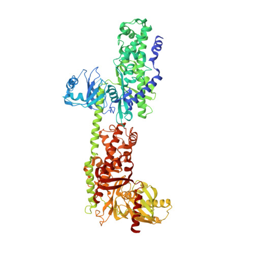
5HG1

Crystal Structure of Human Hexokinase 2 with cmpd 1, a C-2-substituted glucosamine


Experimental Data Snapshot

  • Method: X-RAY DIFFRACTION
  • Resolution: 2.76 Å
  • R-Value Free: 0.266 
  • R-Value Work: 0.218 
  • R-Value Observed: 0.220 

wwPDB Validation   3D Report Full Report


Ligand Structure Quality Assessment 


This is version 1.2 of the entry. See complete history


Literature

Discovery of a Novel 2,6-Disubstituted Glucosamine Series of Potent and Selective Hexokinase 2 Inhibitors.

Lin, H.Zeng, J.Xie, R.Schulz, M.J.Tedesco, R.Qu, J.Erhard, K.F.Mack, J.F.Raha, K.Rendina, A.R.Szewczuk, L.M.Kratz, P.M.Jurewicz, A.J.Cecconie, T.Martens, S.McDevitt, P.J.Martin, J.D.Chen, S.B.Jiang, Y.Nickels, L.Schwartz, B.J.Smallwood, A.Zhao, B.Campobasso, N.Qian, Y.Briand, J.Rominger, C.M.Oleykowski, C.Hardwicke, M.A.Luengo, J.I.

(2016) ACS Med Chem Lett 7: 217-222

  • DOI: https://doi.org/10.1021/acsmedchemlett.5b00214
  • Primary Citation of Related Structures:  
    5HEX, 5HFU, 5HG1

  • PubMed Abstract: 

    A novel series of potent and selective hexokinase 2 (HK2) inhibitors, 2,6-disubstituted glucosamines, has been identified based on HTS hits, exemplified by compound 1. Inhibitor-bound crystal structures revealed that the HK2 enzyme could adopt an "induced-fit" conformation. The SAR study led to the identification of potent HK2 inhibitors, such as compound 34 with greater than 100-fold selectivity over HK1. Compound 25 inhibits in situ glycolysis in a UM-UC-3 bladder tumor cell line via (13)CNMR measurement of [3-(13)C]lactate produced from [1,6-(13)C2]glucose added to the cell culture.


  • Organizational Affiliation

    Cancer Metabolism Chemistry; Cancer Metabolism Biology; and Platform Technology & Sciences, GlaxoSmithKline , 1250 South Collegeville Road, Collegeville, Pennsylvania 19426-0989, United States.


Macromolecules
Find similar proteins by:  (by identity cutoff)  |  3D Structure
Entity ID: 1
MoleculeChains Sequence LengthOrganismDetailsImage
Hexokinase-2922Homo sapiensMutation(s): 0 
Gene Names: HK2
EC: 2.7.1.1
UniProt & NIH Common Fund Data Resources
Find proteins for P52789 (Homo sapiens)
Explore P52789 
Go to UniProtKB:  P52789
PHAROS:  P52789
GTEx:  ENSG00000159399 
Entity Groups  
Sequence Clusters30% Identity50% Identity70% Identity90% Identity95% Identity100% Identity
UniProt GroupP52789
Sequence Annotations
Expand
  • Reference Sequence
Small Molecules
Ligands 3 Unique
IDChains Name / Formula / InChI Key2D Diagram3D Interactions
62C
Query on 62C

Download Ideal Coordinates CCD File 
B [auth A],
C [auth A]
2-deoxy-2-{[(2E)-3-(3,4-dichlorophenyl)prop-2-enoyl]amino}-alpha-D-glucopyranose
C15 H17 Cl2 N O6
ZBHWRKZTEDNOAJ-GJAUZLMESA-N
BG6
Query on BG6

Download Ideal Coordinates CCD File 
D [auth A]6-O-phosphono-beta-D-glucopyranose
C6 H13 O9 P
NBSCHQHZLSJFNQ-VFUOTHLCSA-N
FLC
Query on FLC

Download Ideal Coordinates CCD File 
E [auth A]CITRATE ANION
C6 H5 O7
KRKNYBCHXYNGOX-UHFFFAOYSA-K
Experimental Data & Validation

Experimental Data

  • Method: X-RAY DIFFRACTION
  • Resolution: 2.76 Å
  • R-Value Free: 0.266 
  • R-Value Work: 0.218 
  • R-Value Observed: 0.220 
  • Space Group: P 31 2 1
Unit Cell:
Length ( Å )Angle ( ˚ )
a = 165.12α = 90
b = 165.12β = 90
c = 126.478γ = 120
Software Package:
Software NamePurpose
PHENIXrefinement
XDSdata reduction
Aimlessdata scaling
PHENIXphasing

Structure Validation

View Full Validation Report


Ligand Structure Quality Assessment 


Entry History 

Deposition Data

Revision History  (Full details and data files)

  • Version 1.0: 2016-03-30
    Type: Initial release
  • Version 1.1: 2020-07-29
    Type: Remediation
    Reason: Carbohydrate remediation
    Changes: Data collection, Derived calculations, Structure summary
  • Version 1.2: 2024-03-06
    Changes: Data collection, Database references, Structure summary