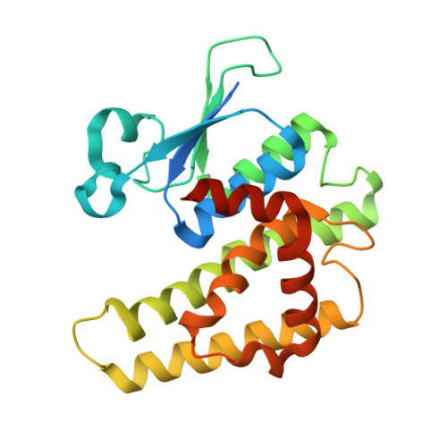
CRYSTAL STRUCTURE OF A GLUTATHIONE S-TRANSFERASE PROTEIN FROM ESCHERICHIA COLI OCh 157:H7 STR. SAKAI (ECs3186, TARGET EFI-507414) WITH BOUND GLUTATHIONE
Himmel, D.M., Toro, R., Al Obaidi, N.F., Morisco, L.L., Wasserman, S.R., Stead, M., Attonito, J.D., Scott Glenn, A., Chamala, S., Chowdhury, S., Lafleur, J., Evans, B., Hillerich, B., Love, J., Seidel, R.D., Whalen, K.L., Gerlt, J.A., Almo, S.C., Enzyme Function Initiative (EFI)To be published.