5HD5 | pdb_00005hd5

Femtosecond Structural Dynamics Drives the Trans/Cis Isomerization in Photoactive Yellow Protein: 200 ns time delay photo-activated (light) structure


Experimental Data Snapshot

  • Method: X-RAY DIFFRACTION
  • Resolution: 1.60 Å
  • R-Value Free: 
    0.374 (Depositor), 0.364 (DCC) 
  • R-Value Work: 
    0.289 (Depositor), 0.284 (DCC) 
  • R-Value Observed: 
    0.293 (Depositor) 

wwPDB Validation 3D Report Full Report

Validation slider image for 5HD5

This is version 1.5 of the entry. See complete history

Literature

Femtosecond structural dynamics drives the trans/cis isomerization in photoactive yellow protein.

Pande, K.Hutchison, C.D.Groenhof, G.Aquila, A.Robinson, J.S.Tenboer, J.Basu, S.Boutet, S.DePonte, D.P.Liang, M.White, T.A.Zatsepin, N.A.Yefanov, O.Morozov, D.Oberthuer, D.Gati, C.Subramanian, G.James, D.Zhao, Y.Koralek, J.Brayshaw, J.Kupitz, C.Conrad, C.Roy-Chowdhury, S.Coe, J.D.Metz, M.Xavier, P.L.Grant, T.D.Koglin, J.E.Ketawala, G.Fromme, R.Srajer, V.Henning, R.Spence, J.C.Ourmazd, A.Schwander, P.Weierstall, U.Frank, M.Fromme, P.Barty, A.Chapman, H.N.Moffat, K.van Thor, J.J.Schmidt, M.

(2016) Science 352: 725-729

  • DOI: https://doi.org/10.1126/science.aad5081
  • Primary Citation Related Structures: 
    5HD3, 5HD5, 5HDC, 5HDD, 5HDS

  • PubMed Abstract: 

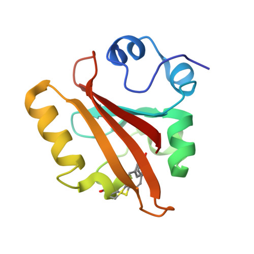
    A variety of organisms have evolved mechanisms to detect and respond to light, in which the response is mediated by protein structural changes after photon absorption. The initial step is often the photoisomerization of a conjugated chromophore. Isomerization occurs on ultrafast time scales and is substantially influenced by the chromophore environment. Here we identify structural changes associated with the earliest steps in the trans-to-cis isomerization of the chromophore in photoactive yellow protein. Femtosecond hard x-ray pulses emitted by the Linac Coherent Light Source were used to conduct time-resolved serial femtosecond crystallography on photoactive yellow protein microcrystals over a time range from 100 femtoseconds to 3 picoseconds to determine the structural dynamics of the photoisomerization reaction.


  • Organizational Affiliation
    • Department of Physics, University of Wisconsin-Milwaukee, Milwaukee, WI 53211, USA. Center for Free Electron Laser Science, Deutsches Elektronen Synchrotron, Notkestrasse 85, 22607 Hamburg, Germany.

Macromolecule Content 

  • Total Structure Weight: 14.03 kDa 
  • Atom Count: 1,244 
  • Modeled Residue Count: 125 
  • Deposited Residue Count: 125 
  • Unique protein chains: 1

Macromolecules

Find similar proteins by:|  3D Structure
Entity ID: 1
MoleculeChains  Sequence LengthOrganismDetailsImage
Photoactive yellow protein125Halorhodospira halophilaMutation(s): 0 
Gene Names: pyp
UniProt
Find proteins for P16113 (Halorhodospira halophila)
Explore P16113 
Go to UniProtKB:  P16113
Entity Groups
Sequence Clusters30% Identity50% Identity70% Identity90% Identity95% Identity100% Identity
UniProt GroupP16113
Sequence Annotations
Expand
Reference Sequence

Small Molecules

Modified Residues  1 Unique
IDChains TypeFormula2D DiagramParent
60F
Query on 60F
A
L-PEPTIDE LINKINGC12 H13 N O4 SCYS

Experimental Data & Validation

Experimental Data

  • Method: X-RAY DIFFRACTION
  • Resolution: 1.60 Å
  • R-Value Free:  0.374 (Depositor), 0.364 (DCC) 
  • R-Value Work:  0.289 (Depositor), 0.284 (DCC) 
  • R-Value Observed: 0.293 (Depositor) 
Space Group: P 63
Unit Cell:
Length ( Å )Angle ( ˚ )
a = 66.9α = 90
b = 66.9β = 90
c = 40.8γ = 120
Software Package:
Software NamePurpose
REFMACrefinement
CrystFELdata reduction
CrystFELdata scaling

Structure Validation

View Full Validation Report


Entry History 

& Funding Information

Deposition Data


Funding OrganizationLocationGrant Number
National Science Foundation (NSF, United States)United StatesNSF-0952643
National Science Foundation (NSF, United States)United StatesNSF-1231306

Revision History  (Full details and data files)

  • Version 1.0: 2016-05-25
    Type: Initial release
  • Version 1.1: 2017-09-13
    Changes: Author supporting evidence, Derived calculations
  • Version 1.2: 2018-02-14
    Changes: Data collection
  • Version 1.3: 2019-11-27
    Changes: Author supporting evidence
  • Version 1.4: 2023-08-16
    Changes: Data collection, Database references
  • Version 1.5: 2023-11-15
    Changes: Data collection, Derived calculations