5HA0

Crystal structure of the LTBP1 leukotriene d4 complex


Experimental Data Snapshot

  • Method: X-RAY DIFFRACTION
  • Resolution: 1.44 Å
  • R-Value Free: 0.188 
  • R-Value Work: 0.160 
  • R-Value Observed: 0.161 

wwPDB Validation   3D Report Full Report


Ligand Structure Quality Assessment 


This is version 1.2 of the entry. See complete history


Literature

Structure and Ligand-Binding Mechanism of a Cysteinyl Leukotriene-Binding Protein from a Blood-Feeding Disease Vector.

Jablonka, W.Pham, V.Nardone, G.Gittis, A.Silva-Cardoso, L.Atella, G.C.Ribeiro, J.M.Andersen, J.F.

(2016) ACS Chem Biol 11: 1934-1944

  • DOI: https://doi.org/10.1021/acschembio.6b00032
  • Primary Citation of Related Structures:  
    5H9K, 5H9L, 5H9N, 5HA0, 5HAE

  • PubMed Abstract: 

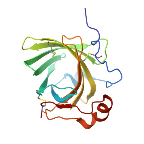
    Blood-feeding disease vectors mitigate the negative effects of hemostasis and inflammation through the binding of small-molecule agonists of these processes by salivary proteins. In this study, a lipocalin protein family member (LTBP1) from the saliva of Rhodnius prolixus, a vector of the pathogen Trypanosoma cruzi, is shown to sequester cysteinyl leukotrienes during feeding to inhibit immediate inflammatory responses. Calorimetric binding experiments showed that LTBP1 binds leukotrienes C4 (LTC4), D4 (LTD4), and E4 (LTE4) but not biogenic amines, adenosine diphosphate, or other eicosanoid compounds. Crystal structures of ligand-free LTBP1 and its complexes with LTC4 and LTD4 reveal a conformational change during binding that brings Tyr114 into close contact with the ligand. LTC4 is cleaved in the complex, leaving free glutathione and a C20 fatty acid. Chromatographic analysis of bound ligands showed only intact LTC4, suggesting that cleavage could be radiation-mediated.


  • Organizational Affiliation

    Laboratory of Malaria and Vector Research, NIAID, National Institutes of Health , Rockville, Maryland 20852, United States.


Macromolecules
Find similar proteins by:  (by identity cutoff)  |  3D Structure
Entity ID: 1
MoleculeChains Sequence LengthOrganismDetailsImage
Lipocalin AI-4156Rhodnius prolixusMutation(s): 0 
UniProt
Find proteins for Q7YT09 (Rhodnius prolixus)
Explore Q7YT09 
Go to UniProtKB:  Q7YT09
Entity Groups  
Sequence Clusters30% Identity50% Identity70% Identity90% Identity95% Identity100% Identity
UniProt GroupQ7YT09
Sequence Annotations
Expand
  • Reference Sequence
Small Molecules
Ligands 1 Unique
IDChains Name / Formula / InChI Key2D Diagram3D Interactions
LTD
Query on LTD

Download Ideal Coordinates CCD File 
B [auth A](5~{S},6~{R},7~{E},9~{E},11~{Z},14~{Z})-6-[(2~{R})-2-azanyl-3-(2-hydroxy-2-oxoethylamino)-3-oxidanylidene-propyl]sulfanyl-5-oxidanyl-icosa-7,9,11,14-tetraenoic acid
C25 H40 N2 O6 S
YEESKJGWJFYOOK-IJHYULJSSA-N
Binding Affinity Annotations 
IDSourceBinding Affinity
LTD Binding MOAD:  5HA0 Kd: 1 (nM) from 1 assay(s)
Experimental Data & Validation

Experimental Data

  • Method: X-RAY DIFFRACTION
  • Resolution: 1.44 Å
  • R-Value Free: 0.188 
  • R-Value Work: 0.160 
  • R-Value Observed: 0.161 
  • Space Group: P 1 21 1
Unit Cell:
Length ( Å )Angle ( ˚ )
a = 34.402α = 90
b = 58.299β = 104.14
c = 36.039γ = 90
Software Package:
Software NamePurpose
PHENIXrefinement
HKL-2000data scaling
PDB_EXTRACTdata extraction
HKL-2000data reduction
PHENIXphasing

Structure Validation

View Full Validation Report


Ligand Structure Quality Assessment 


Entry History 

Deposition Data

Revision History  (Full details and data files)

  • Version 1.0: 2016-05-11
    Type: Initial release
  • Version 1.1: 2016-07-27
    Changes: Database references
  • Version 1.2: 2023-09-27
    Changes: Data collection, Database references, Derived calculations, Refinement description