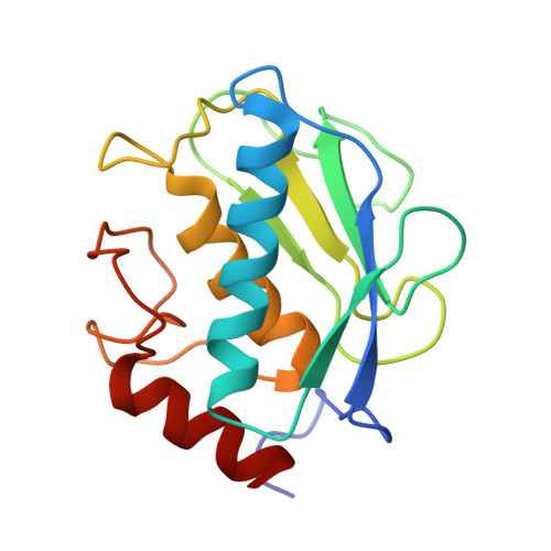
5H8X

Crystal structure of the complex MMP-8/BF471 (catechol inhibitor)


Experimental Data Snapshot

  • Method: X-RAY DIFFRACTION
  • Resolution: 1.30 Å
  • R-Value Free: 0.217 
  • R-Value Work: 0.195 
  • R-Value Observed: 0.196 

Starting Model: experimental
View more details

wwPDB Validation   3D Report Full Report


Ligand Structure Quality Assessment 


This is version 1.2 of the entry. See complete history


Literature

Catechol-based matrix metalloproteinase inhibitors with additional antioxidative activity.

Tauro, M.Laghezza, A.Loiodice, F.Piemontese, L.Caradonna, A.Capelli, D.Montanari, R.Pochetti, G.Di Pizio, A.Agamennone, M.Campestre, C.Tortorella, P.

(2016) J Enzyme Inhib Med Chem 31: 25-37

  • DOI: https://doi.org/10.1080/14756366.2016.1217853
  • Primary Citation of Related Structures:  
    5H8X

  • PubMed Abstract: 

    New catechol-containing chemical entities have been investigated as matrix metalloproteinase inhibitors as well as antioxidant molecules. The combination of the two properties could represent a useful feature due to the potential application in all the pathological processes characterized by increased proteolytic activity and radical oxygen species (ROS) production, such as inflammation and photoaging. A series of catechol-based molecules were synthesized and tested for both proteolytic and oxidative inhibitory activity, and the detailed binding mode was assessed by crystal structure determination of the complex between a catechol derivative and the matrix metalloproteinase-8. Surprisingly, X-ray structure reveals that the catechol oxygens do not coordinates the zinc atom.


  • Organizational Affiliation

    a Department of Tumor Biology , H. Lee Moffitt Cancer Center and Research Institute , Tampa , FL , USA.


Macromolecules
Find similar proteins by:  (by identity cutoff)  |  3D Structure
Entity ID: 1
MoleculeChains Sequence LengthOrganismDetailsImage
Neutrophil collagenase163Homo sapiensMutation(s): 0 
Gene Names: MMP8CLG1
EC: 3.4.24.34
UniProt & NIH Common Fund Data Resources
Find proteins for P22894 (Homo sapiens)
Explore P22894 
Go to UniProtKB:  P22894
PHAROS:  P22894
GTEx:  ENSG00000118113 
Entity Groups  
Sequence Clusters30% Identity50% Identity70% Identity90% Identity95% Identity100% Identity
UniProt GroupP22894
Sequence Annotations
Expand
  • Reference Sequence
Small Molecules
Ligands 5 Unique
IDChains Name / Formula / InChI Key2D Diagram3D Interactions
5XT
Query on 5XT

Download Ideal Coordinates CCD File 
F [auth A]~{N}-[3,4-bis(oxidanyl)phenyl]-4-phenyl-benzenesulfonamide
C18 H15 N O4 S
OGGBNPCRZSBOOJ-UHFFFAOYSA-N
7FY
Query on 7FY

Download Ideal Coordinates CCD File 
G [auth A]~{N}-[4,5-bis(oxidanylidene)cyclohexen-1-yl]-4-phenyl-benzenesulfonamide
C18 H15 N O4 S
APAOLMVDZOWNQC-UHFFFAOYSA-N
MES
Query on MES

Download Ideal Coordinates CCD File 
H [auth A]2-(N-MORPHOLINO)-ETHANESULFONIC ACID
C6 H13 N O4 S
SXGZJKUKBWWHRA-UHFFFAOYSA-N
ZN
Query on ZN

Download Ideal Coordinates CCD File 
D [auth A],
E [auth A]
ZINC ION
Zn
PTFCDOFLOPIGGS-UHFFFAOYSA-N
CA
Query on CA

Download Ideal Coordinates CCD File 
B [auth A],
C [auth A]
CALCIUM ION
Ca
BHPQYMZQTOCNFJ-UHFFFAOYSA-N
Experimental Data & Validation

Experimental Data

  • Method: X-RAY DIFFRACTION
  • Resolution: 1.30 Å
  • R-Value Free: 0.217 
  • R-Value Work: 0.195 
  • R-Value Observed: 0.196 
  • Space Group: P 21 21 21
Unit Cell:
Length ( Å )Angle ( ˚ )
a = 32.91α = 90
b = 68.46β = 90
c = 68.53γ = 90
Software Package:
Software NamePurpose
PHENIXrefinement
iMOSFLMdata reduction
SCALEPACKdata scaling
AMoREphasing

Structure Validation

View Full Validation Report


Ligand Structure Quality Assessment 


Entry History 

Deposition Data

Revision History  (Full details and data files)

  • Version 1.0: 2016-11-02
    Type: Initial release
  • Version 1.1: 2016-12-28
    Changes: Database references
  • Version 1.2: 2024-01-10
    Changes: Data collection, Database references, Derived calculations, Refinement description