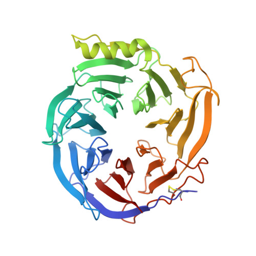
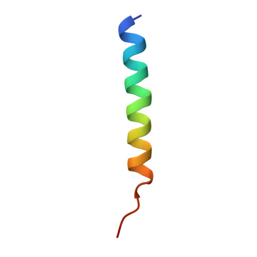
5H17

EED in complex with PRC2 allosteric inhibitor EED210


Experimental Data Snapshot

  • Method: X-RAY DIFFRACTION
  • Resolution: 2.30 Å
  • R-Value Free: 0.239 
  • R-Value Work: 0.189 
  • R-Value Observed: 0.192 

Starting Model: experimental
View more details

wwPDB Validation   3D Report Full Report


Ligand Structure Quality Assessment 


This is version 1.2 of the entry. See complete history


Literature

Discovery and Molecular Basis of a Diverse Set of Polycomb Repressive Complex 2 Inhibitors Recognition by EED

Li, L.Zhang, H.Zhang, M.Zhao, M.Feng, L.Luo, X.Gao, Z.Huang, Y.Ardayfio, O.Zhang, J.H.Lin, Y.Fan, H.Mi, Y.Li, G.Liu, L.Feng, L.Luo, F.Teng, L.Qi, W.Ottl, J.Lingel, A.Bussiere, D.E.Yu, Z.Atadja, P.Lu, C.Li, E.Gu, J.Zhao, K.

(2017) PLoS One 12: e0169855-e0169855

  • DOI: https://doi.org/10.1371/journal.pone.0169855
  • Primary Citation of Related Structures:  
    5H13, 5H14, 5H15, 5H17, 5H19

  • PubMed Abstract: 

    Polycomb repressive complex 2 (PRC2), a histone H3 lysine 27 methyltransferase, plays a key role in gene regulation and is a known epigenetics drug target for cancer therapy. The WD40 domain-containing protein EED is the regulatory subunit of PRC2. It binds to the tri-methylated lysine 27 of the histone H3 (H3K27me3), and through which stimulates the activity of PRC2 allosterically. Recently, we disclosed a novel PRC2 inhibitor EED226 which binds to the K27me3-pocket on EED and showed strong antitumor activity in xenograft mice model. Here, we further report the identification and validation of four other EED binders along with EED162, the parental compound of EED226. The crystal structures for all these five compounds in complex with EED revealed a common deep pocket induced by the binding of this diverse set of compounds. This pocket was created after significant conformational rearrangement of the aromatic cage residues (Y365, Y148 and F97) in the H3K27me3 binding pocket of EED, the width of which was delineated by the side chains of these rearranged residues. In addition, all five compounds interact with the Arg367 at the bottom of the pocket. Each compound also displays unique features in its interaction with EED, suggesting the dynamics of the H3K27me3 pocket in accommodating the binding of different compounds. Our results provide structural insights for rational design of novel EED binder for the inhibition of PRC2 complex activity.


  • Organizational Affiliation

    China Novartis Institutes for BioMedical Research, Shanghai, China.


Macromolecules
Find similar proteins by:  (by identity cutoff)  |  3D Structure
Entity ID: 1
MoleculeChains Sequence LengthOrganismDetailsImage
Polycomb protein EED367Homo sapiensMutation(s): 0 
Gene Names: EED
UniProt & NIH Common Fund Data Resources
Find proteins for O75530 (Homo sapiens)
Explore O75530 
Go to UniProtKB:  O75530
PHAROS:  O75530
GTEx:  ENSG00000074266 
Entity Groups  
Sequence Clusters30% Identity50% Identity70% Identity90% Identity95% Identity100% Identity
UniProt GroupO75530
Sequence Annotations
Expand
  • Reference Sequence
Find similar proteins by:  (by identity cutoff)  |  3D Structure
Entity ID: 2
MoleculeChains Sequence LengthOrganismDetailsImage
Histone-lysine N-methyltransferase EZH229Homo sapiensMutation(s): 0 
EC: 2.1.1.43 (PDB Primary Data), 2.1.1.356 (UniProt)
UniProt & NIH Common Fund Data Resources
Find proteins for Q15910 (Homo sapiens)
Explore Q15910 
Go to UniProtKB:  Q15910
PHAROS:  Q15910
GTEx:  ENSG00000106462 
Entity Groups  
Sequence Clusters30% Identity50% Identity70% Identity90% Identity95% Identity100% Identity
UniProt GroupQ15910
Sequence Annotations
Expand
  • Reference Sequence
Small Molecules
Ligands 1 Unique
IDChains Name / Formula / InChI Key2D Diagram3D Interactions
LQE
Query on LQE

Download Ideal Coordinates CCD File 
C [auth A](3R,4aS,10aS)-6-methoxy-3-[(3-methoxyphenyl)methyl]-1-methyl-3,4,4a,5,10,10a-hexahydro-2H-benzo[g]quinoline
C23 H29 N O2
QZUPQRHYHMMHGC-JLMWRMLUSA-N
Binding Affinity Annotations 
IDSourceBinding Affinity
LQE BindingDB:  5H17 IC50: min: 890, max: 2500 (nM) from 2 assay(s)
Experimental Data & Validation

Experimental Data

  • Method: X-RAY DIFFRACTION
  • Resolution: 2.30 Å
  • R-Value Free: 0.239 
  • R-Value Work: 0.189 
  • R-Value Observed: 0.192 
  • Space Group: C 2 2 21
Unit Cell:
Length ( Å )Angle ( ˚ )
a = 50.56α = 90
b = 92.28β = 90
c = 180.48γ = 90
Software Package:
Software NamePurpose
XSCALEdata scaling
BUSTERrefinement
PDB_EXTRACTdata extraction
MOLREPphasing

Structure Validation

View Full Validation Report


Ligand Structure Quality Assessment 


Entry History 

Deposition Data

Revision History  (Full details and data files)

  • Version 1.0: 2017-01-25
    Type: Initial release
  • Version 1.1: 2023-11-08
    Changes: Data collection, Database references, Refinement description
  • Version 1.2: 2024-11-20
    Changes: Structure summary