5GZT

Crystal Structure of Chitinase ChiW from Paenibacillus sp. str. FPU-7 Reveals a Novel Type of Bacterial Cell-Surface-Expressed Multi-Modular Enzyme Machinery


Experimental Data Snapshot

  • Method: X-RAY DIFFRACTION
  • Resolution: 2.10 Å
  • R-Value Free: 0.221 
  • R-Value Work: 0.175 
  • R-Value Observed: 0.178 

Starting Model: experimental
View more details

wwPDB Validation   3D Report Full Report


This is version 1.3 of the entry. See complete history


Literature

Crystal Structure of Chitinase ChiW from Paenibacillus sp. str. FPU-7 Reveals a Novel Type of Bacterial Cell-Surface-Expressed Multi-Modular Enzyme Machinery

Itoh, T.Hibi, T.Suzuki, F.Sugimoto, I.Fujiwara, A.Inaka, K.Tanaka, H.Ohta, K.Fujii, Y.Taketo, A.Kimoto, H.

(2016) PLoS One 11: e0167310-e0167310

  • DOI: https://doi.org/10.1371/journal.pone.0167310
  • Primary Citation of Related Structures:  
    5GZT, 5GZU, 5GZV

  • PubMed Abstract: 

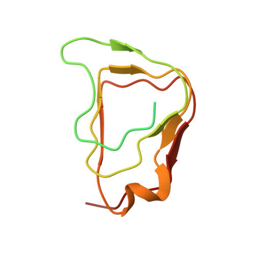
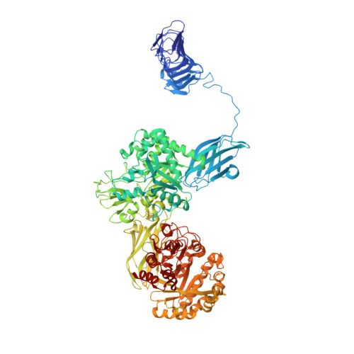
    The Gram-positive bacterium Paenibacillus sp. str. FPU-7 effectively hydrolyzes chitin by using a number of chitinases. A unique chitinase with two catalytic domains, ChiW, is expressed on the cell surface of this bacterium and has high activity towards various chitins, even crystalline chitin. Here, the crystal structure of ChiW at 2.1 Å resolution is presented and describes how the enzyme degrades chitin on the bacterial cell surface. The crystal structure revealed a unique multi-modular architecture composed of six domains to function efficiently on the cell surface: a right-handed β-helix domain (carbohydrate-binding module family 54, CBM-54), a Gly-Ser-rich loop, 1st immunoglobulin-like (Ig-like) fold domain, 1st β/α-barrel catalytic domain (glycoside hydrolase family 18, GH-18), 2nd Ig-like fold domain and 2nd β/α-barrel catalytic domain (GH-18). The structure of the CBM-54, flexibly linked to the catalytic region of ChiW, is described here for the first time. It is similar to those of carbohydrate lyases but displayed no detectable carbohydrate degradation activities. The CBM-54 of ChiW bound to cell wall polysaccharides, such as chin, chitosan, β-1,3-glucan, xylan and cellulose. The structural and biochemical data obtained here also indicated that the enzyme has deep and short active site clefts with endo-acting character. The affinity of CBM-54 towards cell wall polysaccharides and the degradation pattern of the catalytic domains may help to efficiently decompose the cell wall chitin through the contact surface. Furthermore, we clarify that other Gram-positive bacteria possess similar cell-surface-expressed multi-modular enzymes for cell wall polysaccharide degradation.


  • Organizational Affiliation

    Department of Bioscience, Fukui Prefectural University, Yoshida-gun, Fukui, Japan.


Macromolecules
Find similar proteins by:  (by identity cutoff)  |  3D Structure
Entity ID: 1
MoleculeChains Sequence LengthOrganismDetailsImage
Chitinase108Paenibacillus sp. FPU-7Mutation(s): 0 
Gene Names: chiW
UniProt
Find proteins for K7ZLW6 (Paenibacillus sp. FPU-7)
Explore K7ZLW6 
Go to UniProtKB:  K7ZLW6
Entity Groups  
Sequence Clusters30% Identity50% Identity70% Identity90% Identity95% Identity100% Identity
UniProt GroupK7ZLW6
Sequence Annotations
Expand
  • Reference Sequence
Find similar proteins by:  (by identity cutoff)  |  3D Structure
Entity ID: 2
MoleculeChains Sequence LengthOrganismDetailsImage
Chitinase1,136Paenibacillus sp. FPU-7Mutation(s): 0 
Gene Names: chiW
UniProt
Find proteins for K7ZLW6 (Paenibacillus sp. FPU-7)
Explore K7ZLW6 
Go to UniProtKB:  K7ZLW6
Entity Groups  
Sequence Clusters30% Identity50% Identity70% Identity90% Identity95% Identity100% Identity
UniProt GroupK7ZLW6
Sequence Annotations
Expand
  • Reference Sequence
Small Molecules
Ligands 3 Unique
IDChains Name / Formula / InChI Key2D Diagram3D Interactions
PO4
Query on PO4

Download Ideal Coordinates CCD File 
D [auth B]PHOSPHATE ION
O4 P
NBIIXXVUZAFLBC-UHFFFAOYSA-K
FMT
Query on FMT

Download Ideal Coordinates CCD File 
E [auth B]
F [auth B]
G [auth B]
H [auth B]
I [auth B]
E [auth B],
F [auth B],
G [auth B],
H [auth B],
I [auth B],
J [auth B],
K [auth B],
L [auth B],
M [auth B],
N [auth B]
FORMIC ACID
C H2 O2
BDAGIHXWWSANSR-UHFFFAOYSA-N
NA
Query on NA

Download Ideal Coordinates CCD File 
C [auth B]SODIUM ION
Na
FKNQFGJONOIPTF-UHFFFAOYSA-N
Experimental Data & Validation

Experimental Data

  • Method: X-RAY DIFFRACTION
  • Resolution: 2.10 Å
  • R-Value Free: 0.221 
  • R-Value Work: 0.175 
  • R-Value Observed: 0.178 
  • Space Group: P 21 21 21
Unit Cell:
Length ( Å )Angle ( ˚ )
a = 114.339α = 90
b = 123.486β = 90
c = 130.856γ = 90
Software Package:
Software NamePurpose
REFMACrefinement
HKL-2000data reduction
HKL-2000data scaling
PHASERphasing

Structure Validation

View Full Validation Report


Entry History 

Deposition Data

Revision History  (Full details and data files)

  • Version 1.0: 2016-12-28
    Type: Initial release
  • Version 1.1: 2017-01-04
    Changes: Derived calculations
  • Version 1.2: 2020-02-26
    Changes: Data collection
  • Version 1.3: 2023-11-08
    Changes: Data collection, Database references, Refinement description