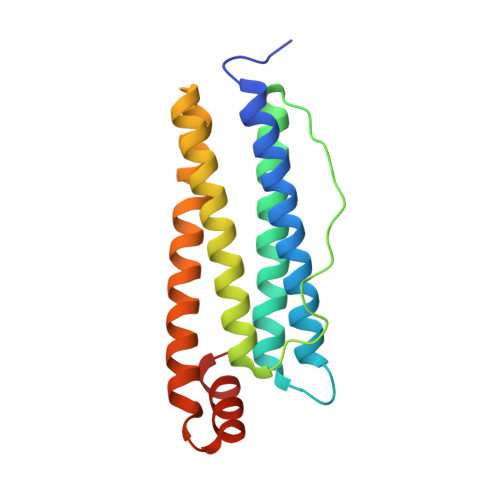
5GU3

Crystal structure of Au(E).CL-apo-E45C/R52C-rHLFr


Experimental Data Snapshot

  • Method: X-RAY DIFFRACTION
  • Resolution: 2.03 Å
  • R-Value Free: 0.244 
  • R-Value Work: 0.189 
  • R-Value Observed: 0.192 

Starting Model: experimental
View more details

wwPDB Validation   3D Report Full Report


This is version 1.2 of the entry. See complete history


Literature

Observation of gold sub-nanocluster nucleation within a crystalline protein cage

Maity, B.Abe, S.Ueno, T.

(2017) Nat Commun 8: 14820-14820

  • DOI: https://doi.org/10.1038/ncomms14820
  • Primary Citation of Related Structures:  
    5GU0, 5GU1, 5GU2, 5GU3

  • PubMed Abstract: 

    Protein scaffolds provide unique metal coordination environments that promote biomineralization processes. It is expected that protein scaffolds can be developed to prepare inorganic nanomaterials with important biomedical and material applications. Despite many promising applications, it remains challenging to elucidate the detailed mechanisms of formation of metal nanoparticles in protein environments. In the present work, we describe a crystalline protein cage constructed by crosslinking treatment of a single crystal of apo-ferritin for structural characterization of the formation of sub-nanocluster with reduction reaction. The crystal structure analysis shows the gradual movement of the Au ions towards the centre of the three-fold symmetric channels of the protein cage to form a sub-nanocluster with accompanying significant conformational changes of the amino-acid residues bound to Au ions during the process. These results contribute to our understanding of metal core formation as well as interactions of the metal core with the protein environment.


  • Organizational Affiliation

    School of Life Science and Technology, Tokyo Institute of Technology, 4259-B55, Nagatsuta-cho, Midori-ku, Yokohama 226-8501, Japan.


Macromolecules
Find similar proteins by:  (by identity cutoff)  |  3D Structure
Entity ID: 1
MoleculeChains Sequence LengthOrganismDetailsImage
Ferritin light chainA [auth X]174Equus caballusMutation(s): 2 
Gene Names: FTL
UniProt
Find proteins for P02791 (Equus caballus)
Explore P02791 
Go to UniProtKB:  P02791
Entity Groups  
Sequence Clusters30% Identity50% Identity70% Identity90% Identity95% Identity100% Identity
UniProt GroupP02791
Sequence Annotations
Expand
  • Reference Sequence
Small Molecules
Ligands 4 Unique
IDChains Name / Formula / InChI Key2D Diagram3D Interactions
AU
Query on AU

Download Ideal Coordinates CCD File 
D [auth X]
E [auth X]
F [auth X]
G [auth X]
H [auth X]
D [auth X],
E [auth X],
F [auth X],
G [auth X],
H [auth X],
I [auth X]
GOLD ION
Au
ZBKIUFWVEIBQRT-UHFFFAOYSA-N
CD
Query on CD

Download Ideal Coordinates CCD File 
J [auth X],
K [auth X],
L [auth X],
M [auth X],
N [auth X]
CADMIUM ION
Cd
WLZRMCYVCSSEQC-UHFFFAOYSA-N
SO4
Query on SO4

Download Ideal Coordinates CCD File 
B [auth X],
C [auth X]
SULFATE ION
O4 S
QAOWNCQODCNURD-UHFFFAOYSA-L
EDO
Query on EDO

Download Ideal Coordinates CCD File 
O [auth X],
P [auth X],
Q [auth X]
1,2-ETHANEDIOL
C2 H6 O2
LYCAIKOWRPUZTN-UHFFFAOYSA-N
Experimental Data & Validation

Experimental Data

  • Method: X-RAY DIFFRACTION
  • Resolution: 2.03 Å
  • R-Value Free: 0.244 
  • R-Value Work: 0.189 
  • R-Value Observed: 0.192 
  • Space Group: F 4 3 2
Unit Cell:
Length ( Å )Angle ( ˚ )
a = 181.502α = 90
b = 181.502β = 90
c = 181.502γ = 90
Software Package:
Software NamePurpose
REFMACrefinement
HKL-2000data scaling
HKL-2000data processing

Structure Validation

View Full Validation Report


Entry History 

Deposition Data

Revision History  (Full details and data files)

  • Version 1.0: 2017-03-29
    Type: Initial release
  • Version 1.1: 2020-02-26
    Changes: Data collection
  • Version 1.2: 2023-11-08
    Changes: Data collection, Database references, Derived calculations, Refinement description