


3D Crystal Structure of LsrB Bound to Furanosyl diester (R)-THMF, from Salmonella typhi
Gopinath, S., Perumal, P., Rahul, R., Arockiasamy, A., Sundarabaalaji, N.To be published.
Experimental Data Snapshot
Starting Model: experimental
View more details
Entity ID: 1 | |||||
|---|---|---|---|---|---|
| Molecule | Chains | Sequence Length | Organism | Details | Image |
| Autoinducer 2-binding protein LsrB | 340 | Salmonella enterica subsp. enterica serovar Typhi | Mutation(s): 0 Gene Names: lsrB, STY3793, t3541 | 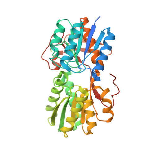 | |
UniProt | |||||
Entity Groups | |||||
| Sequence Clusters | 30% Identity50% Identity70% Identity90% Identity95% Identity100% Identity | ||||
| UniProt Group | Q8Z2X8 | ||||
Sequence AnnotationsExpand | |||||
Reference Sequence | |||||
| Ligands 1 Unique | |||||
|---|---|---|---|---|---|
| ID | Chains | Name / Formula / InChI Key | 2D Diagram | 3D Interactions | |
| PAV Download:Ideal Coordinates CCD File | B [auth A] | (2R,4S)-2-methyl-2,3,3,4-tetrahydroxytetrahydrofuran C5 H10 O5 BVIYGXUQVXBHQS-IUYQGCFVSA-N |  | ||
| Length ( Å ) | Angle ( ˚ ) |
|---|---|
| a = 41.582 | α = 90 |
| b = 61.754 | β = 90 |
| c = 115.753 | γ = 90 |
| Software Name | Purpose |
|---|---|
| PHENIX | refinement |
| HKL-2000 | data processing |
| HKL-2000 | data scaling |
| PHENIX | phasing |
| PHENIX | model building |
| Funding Organization | Location | Grant Number |
|---|---|---|
| Indian Council of Medical Research | India | 2011-15260 |


