5GPG | pdb_00005gpg

Co-crystal structure of the FK506 binding domain of human FKBP25, Rapamycin and the FRB domain of human mTOR


Experimental Data Snapshot

  • Method: X-RAY DIFFRACTION
  • Resolution: 1.67 Å
  • R-Value Free: 
    0.212 (Depositor), 0.216 (DCC) 
  • R-Value Work: 
    0.174 (Depositor), 0.179 (DCC) 
  • R-Value Observed: 
    0.176 (Depositor) 

Starting Models: experimental
View more details

wwPDB Validation 3D Report Full Report

Validation slider image for 5GPG

Ligand Structure Quality Assessment 


This is version 1.2 of the entry. See complete history

Literature

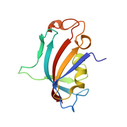
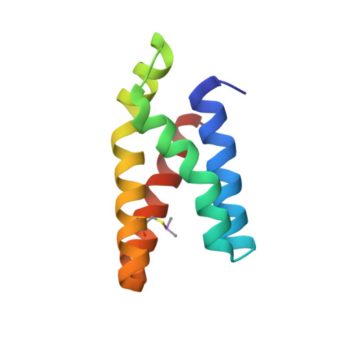
Proximity-Directed Labeling Reveals a New Rapamycin-Induced Heterodimer of FKBP25 and FRB in Live Cells

Lee, S.Y.Lee, H.Lee, H.K.Lee, S.W.Ha, S.C.Kwon, T.Seo, J.K.Lee, C.Rhee, H.W.

(2016) ACS Cent Sci 2: 506-516

  • DOI: https://doi.org/10.1021/acscentsci.6b00137
  • Primary Citation Related Structures: 
    5GPG

  • PubMed Abstract: 

    Mammalian target of rapamycin (mTOR) signaling is a core pathway in cellular metabolism, and control of the mTOR pathway by rapamycin shows potential for the treatment of metabolic diseases. In this study, we employed a new proximity biotin-labeling method using promiscuous biotin ligase (pBirA) to identify unknown elements in the rapamycin-induced interactome on the FK506-rapamycin binding (FRB) domain in living cells. FKBP25 showed the strongest biotin labeling by FRB-pBirA in the presence of rapamycin. Immunoprecipitation and immunofluorescence experiments confirmed that endogenous FKBP25 has a rapamycin-induced physical interaction with the FRB domain. Furthermore, the crystal structure of the ternary complex of FRB-rapamycin-FKBP25 was determined at 1.67-Å resolution. In this crystal structure we found that the conformational changes of FRB generate a hole where there is a methionine-rich space, and covalent metalloid coordination was observed at C2085 of FRB located at the bottom of the hole. Our results imply that FKBP25 might have a unique physiological role related to metallomics in mTOR signaling.


  • Organizational Affiliation
    • Department of Chemistry, Department of Biological Sciences, UNIST Central Research Facilities (UCRF), and Deparment of Biomedical Engineering, Ulsan National Institute of Science and Technology (UNIST) , Ulsan 44919, Korea.

Macromolecule Content 

  • Total Structure Weight: 25.31 kDa 
  • Atom Count: 2,009 
  • Modeled Residue Count: 203 
  • Deposited Residue Count: 210 
  • Unique protein chains: 2

Macromolecules

Find similar proteins by:|  3D Structure
Entity ID: 1
MoleculeChains  Sequence LengthOrganismDetailsImage
Peptidyl-prolyl cis-trans isomerase FKBP3117Homo sapiensMutation(s): 0 
Gene Names: FKBP3FKBP25
EC: 5.2.1.8
UniProt & NIH Common Fund Data Resources
Find proteins for Q00688 (Homo sapiens)
Explore Q00688 
Go to UniProtKB:  Q00688
PHAROS:  Q00688
GTEx:  ENSG00000100442 
Entity Groups
Sequence Clusters30% Identity50% Identity70% Identity90% Identity95% Identity100% Identity
UniProt GroupQ00688
Sequence Annotations
Expand
Reference Sequence
Find similar proteins by:|  3D Structure
Entity ID: 2
MoleculeChains  Sequence LengthOrganismDetailsImage
Serine/threonine-protein kinase mTOR93Homo sapiensMutation(s): 0 
Gene Names: MTORFRAPFRAP1FRAP2RAFT1RAPT1
EC: 2.7.11.1 (PDB Primary Data), 2.7.10.2 (UniProt)
UniProt & NIH Common Fund Data Resources
Find proteins for P42345 (Homo sapiens)
Explore P42345 
Go to UniProtKB:  P42345
PHAROS:  P42345
GTEx:  ENSG00000198793 
Entity Groups
Sequence Clusters30% Identity50% Identity70% Identity90% Identity95% Identity100% Identity
UniProt GroupP42345
Sequence Annotations
Expand
Reference Sequence

Small Molecules

Ligands 1 Unique
IDChains Name / Formula / InChI Key2D Diagram3D Interactions
RAP

Query on RAP


Download:Ideal Coordinates CCD File
C [auth A]RAPAMYCIN IMMUNOSUPPRESSANT DRUG
C51 H79 N O13
QFJCIRLUMZQUOT-HPLJOQBZSA-N
Modified Residues  1 Unique
IDChains TypeFormula2D DiagramParent
CAS
Query on CAS
B
L-PEPTIDE LINKINGC5 H12 As N O2 SCYS

Experimental Data & Validation

Experimental Data

  • Method: X-RAY DIFFRACTION
  • Resolution: 1.67 Å
  • R-Value Free:  0.212 (Depositor), 0.216 (DCC) 
  • R-Value Work:  0.174 (Depositor), 0.179 (DCC) 
  • R-Value Observed: 0.176 (Depositor) 
Space Group: P 21 21 21
Unit Cell:
Length ( Å )Angle ( ˚ )
a = 46.038α = 90
b = 58.352β = 90
c = 86.502γ = 90
Software Package:
Software NamePurpose
PHENIXrefinement
HKL-2000data processing
Cootmodel building
PHENIXphasing
HKL-2000data reduction
HKL-2000data scaling

Structure Validation

View Full Validation Report


Ligand Structure Quality Assessment 


Entry History 

Deposition Data

Revision History  (Full details and data files)

  • Version 1.0: 2016-10-12
    Type: Initial release
  • Version 1.1: 2023-11-08
    Changes: Data collection, Database references, Derived calculations, Refinement description
  • Version 1.2: 2024-11-13
    Changes: Structure summary