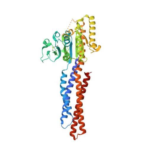
RCSB PDB - 5GOE: Truncated mitofusin-1, GDP-bound

 5GOE

Truncated mitofusin-1, GDP-bound

  • Classification: HYDROLASE
  • Organism(s): Homo sapiens
  • Expression System: Escherichia coli
  • Mutation(s): No 

  • Deposited: 2016-07-26 Released: 2017-01-25 
  • Deposition Author(s): Cao, Y.L., Gao, S.
  • Funding Organization(s): Ministry of Science and Technology of the People's Republic of China, National Natural Science Foundation of China, Ministry of Education of the People's Republic of China

Experimental Data Snapshot

  • Method: X-RAY DIFFRACTION
  • Resolution: 1.80 Å
  • R-Value Free: 0.204 
  • R-Value Work: 0.177 
  • R-Value Observed: 0.178 

wwPDB Validation   3D Report Full Report


Ligand Structure Quality Assessment 

Created with Raphaël 2.3.0Worse 01 BetterLigand structure goodness of fit to experimental dataBest fitted GDPClick on this verticalbar to view details

This is version 1.3 of the entry. See complete history


Literature

MFN1 structures reveal nucleotide-triggered dimerization critical for mitochondrial fusion

Cao, Y.L.Meng, S.Chen, Y.Feng, J.X.Gu, D.D.Yu, B.Li, Y.J.Yang, J.Y.Liao, S.Chan, D.C.Gao, S.

(2017) Nature 542: 372-376

  • DOI: https://doi.org/10.1038/nature21077
  • Primary Citation of Related Structures:  
    5GO4, 5GOE, 5GOF, 5GOM

  • PubMed Abstract: 

    Mitochondria are double-membraned organelles with variable shapes influenced by metabolic conditions, developmental stage, and environmental stimuli. Their dynamic morphology is a result of regulated and balanced fusion and fission processes. Fusion is crucial for the health and physiological functions of mitochondria, including complementation of damaged mitochondrial DNAs and the maintenance of membrane potential. Mitofusins are dynamin-related GTPases that are essential for mitochondrial fusion. They are embedded in the mitochondrial outer membrane and thought to fuse adjacent mitochondria via combined oligomerization and GTP hydrolysis. However, the molecular mechanisms of this process remain unknown. Here we present crystal structures of engineered human MFN1 containing the GTPase domain and a helical domain during different stages of GTP hydrolysis. The helical domain is composed of elements from widely dispersed sequence regions of MFN1 and resembles the 'neck' of the bacterial dynamin-like protein. The structures reveal unique features of its catalytic machinery and explain how GTP binding induces conformational changes to promote GTPase domain dimerization in the transition state. Disruption of GTPase domain dimerization abolishes the fusogenic activity of MFN1. Moreover, a conserved aspartate residue trigger was found to affect mitochondrial elongation in MFN1, probably through a GTP-loading-dependent domain rearrangement. Thus, we propose a mechanistic model for MFN1-mediated mitochondrial tethering, and our results shed light on the molecular basis of mitochondrial fusion and mitofusin-related human neuromuscular disorders.


  • Organizational Affiliation

    State Key Laboratory of Oncology in South China, Collaborative Innovation Center for Cancer Medicine, Sun Yat-sen University Cancer Center, Guangzhou 510060, China.


Macromolecules
Find similar proteins by:  (by identity cutoff)  |  3D Structure
Entity ID: 1
MoleculeChains Sequence LengthOrganismDetailsImage
Mitofusin-1422Homo sapiensMutation(s): 0 
Gene Names: MFN1
EC: 3.6.5
UniProt & NIH Common Fund Data Resources
Find proteins for Q8IWA4 (Homo sapiens)
Explore Q8IWA4 
Go to UniProtKB:  Q8IWA4
PHAROS:  Q8IWA4
GTEx:  ENSG00000171109 
Entity Groups  
Sequence Clusters30% Identity50% Identity70% Identity90% Identity95% Identity100% Identity
UniProt GroupQ8IWA4
Sequence Annotations
Expand
  • Reference Sequence
Small Molecules
Ligands 1 Unique
IDChains Name / Formula / InChI Key2D Diagram3D Interactions
GDP
Query on GDP

Download Ideal Coordinates CCD File 
B [auth A]GUANOSINE-5'-DIPHOSPHATE
C10 H15 N5 O11 P2
QGWNDRXFNXRZMB-UUOKFMHZSA-N
Experimental Data & Validation

Experimental Data

  • Method: X-RAY DIFFRACTION
  • Resolution: 1.80 Å
  • R-Value Free: 0.204 
  • R-Value Work: 0.177 
  • R-Value Observed: 0.178 
  • Space Group: P 21 21 21
Unit Cell:
Length ( Å )Angle ( ˚ )
a = 70.42α = 90
b = 72.91β = 90
c = 94.97γ = 90
Software Package:
Software NamePurpose
PHENIXrefinement
XDSdata reduction
XDSdata scaling
MOLREPphasing

Structure Validation

View Full Validation Report


Ligand Structure Quality Assessment 

Created with Raphaël 2.3.0Worse 01 BetterLigand structure goodness of fit to experimental dataBest fitted GDPClick on this verticalbar to view details

Entry History & Funding Information

Deposition Data


Funding OrganizationLocationGrant Number
Ministry of Science and Technology of the People's Republic of ChinaChina2013CB910500
National Natural Science Foundation of ChinaChina31200553
Ministry of Education of the People's Republic of ChinaChinaNCET-12-0567

Revision History  (Full details and data files)

  • Version 1.0: 2017-01-25
    Type: Initial release
  • Version 1.1: 2017-02-01
    Changes: Structure summary
  • Version 1.2: 2017-03-08
    Changes: Database references
  • Version 1.3: 2024-10-23
    Changes: Advisory, Data collection, Database references, Structure summary