


The structure of Vibrio anguillarum775 AngB-ICL protein
Du, J.S., Deng, T., Ma, Q.G.To be published.
Experimental Data Snapshot
Starting Model: experimental
View more details
wwPDB Validation 3D Report Full Report

Entity ID: 1 | |||||
|---|---|---|---|---|---|
| Molecule | Chains | Sequence Length | Organism | Details | Image |
| Ischorismate lyase | 208 | Vibrio anguillarum 775 | Mutation(s): 0 Gene Names: G, angB | 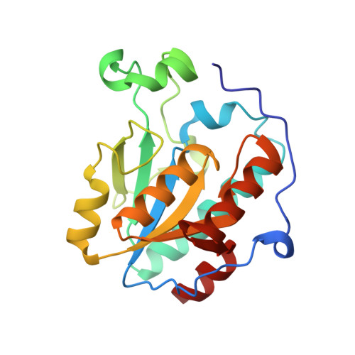 | |
Entity ID: 2 | |||||
|---|---|---|---|---|---|
| Molecule | Chains | Sequence Length | Organism | Details | Image |
| Ischorismate lyase | 208 | Vibrio anguillarum 775 | Mutation(s): 1 Gene Names: G, angB | 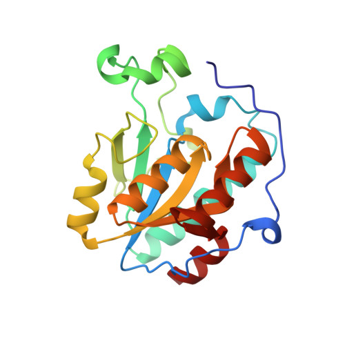 | |
| Length ( Å ) | Angle ( ˚ ) |
|---|---|
| a = 140.19 | α = 90 |
| b = 140.19 | β = 90 |
| c = 59.3 | γ = 120 |
| Software Name | Purpose |
|---|---|
| PHENIX | refinement |
| HKL-2000 | data processing |
| PHENIX | refinement |


