5GKF | pdb_00005gkf

Structure of EndoMS-dsDNA1' complex


Experimental Data Snapshot

  • Method: X-RAY DIFFRACTION
  • Resolution: 2.80 Å
  • R-Value Free: 
    0.247 (Depositor), 0.251 (DCC) 
  • R-Value Work: 
    0.171 (Depositor), 0.183 (DCC) 
  • R-Value Observed: 
    0.175 (Depositor) 

Starting Model: experimental
View more details

wwPDB Validation 3D Report Full Report

Validation slider image for 5GKF

This is version 1.3 of the entry. See complete history

Literature

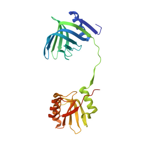
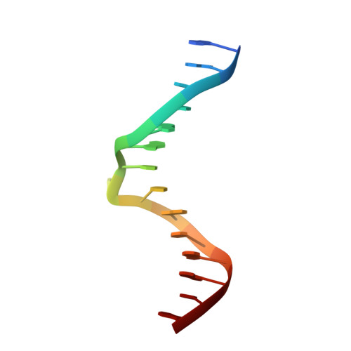
Structure of the EndoMS-DNA Complex as Mismatch Restriction Endonuclease

Nakae, S.Hijikata, A.Tsuji, T.Yonezawa, K.Kouyama, K.I.Mayanagi, K.Ishino, S.Ishino, Y.Shirai, T.

(2016) Structure 24: 1960-1971

  • DOI: https://doi.org/10.1016/j.str.2016.09.005
  • Primary Citation Related Structures: 
    5GKE, 5GKF, 5GKG, 5GKH, 5GKI, 5GKJ

  • PubMed Abstract: 

    Archaeal NucS nuclease was thought to degrade the single-stranded region of branched DNA, which contains flapped and splayed DNA. However, recent findings indicated that EndoMS, the orthologous enzyme of NucS, specifically cleaves double-stranded DNA (dsDNA) containing mismatched bases. In this study, we determined the structure of the EndoMS-DNA complex. The complex structure of the EndoMS dimer with dsDNA unexpectedly revealed that the mismatched bases were flipped out into binding sites, and the overall architecture most resembled that of restriction enzymes. The structure of the apo form was similar to the reported structure of Pyrococcus abyssi NucS, indicating that movement of the C-terminal domain from the resting state was required for activity. In addition, a model of the EndoMS-PCNA-DNA complex was preliminarily verified with electron microscopy. The structures strongly support the idea that EndoMS acts in a mismatch repair pathway.


  • Organizational Affiliation
    • Department of Bioscience, Nagahama Institute of Bio-Science and Technology, Tamura 1266, Nagahama, Shiga 526-0829, Japan.

Macromolecule Content 

  • Total Structure Weight: 67.01 kDa 
  • Atom Count: 5,093 
  • Modeled Residue Count: 508 
  • Deposited Residue Count: 534 
  • Unique protein chains: 1
  • Unique nucleic acid chains: 2

Macromolecules


Find similar proteins by:|  3D Structure
Entity ID: 1
MoleculeChains  Sequence LengthOrganismDetailsImage
Endonuclease EndoMS
A, B
252Thermococcus kodakarensis KOD1Mutation(s): 1 
Gene Names: TK1898
EC: 3.1
UniProt
Find proteins for Q5JER9 (Thermococcus kodakarensis (strain ATCC BAA-918 / JCM 12380 / KOD1))
Explore Q5JER9 
Go to UniProtKB:  Q5JER9
Entity Groups
Sequence Clusters30% Identity50% Identity70% Identity90% Identity95% Identity100% Identity
UniProt GroupQ5JER9
Sequence Annotations
Expand
Reference Sequence
Find similar nucleic acids by:  Sequence
Entity ID: 2
MoleculeChains LengthOrganismImage
DNA (5'-D(*CP*GP*CP*TP*AP*CP*AP*TP*GP*TP*CP*GP*TP*CP*C)-3')15synthetic construct
Sequence Annotations
Expand
Reference Sequence
Find similar nucleic acids by:  Sequence
Entity ID: 3
MoleculeChains LengthOrganismImage
DNA (5'-D(*GP*GP*AP*CP*GP*AP*CP*TP*TP*GP*TP*AP*GP*CP*G)-3')15synthetic construct
Sequence Annotations
Expand
Reference Sequence

Experimental Data & Validation

Experimental Data

  • Method: X-RAY DIFFRACTION
  • Resolution: 2.80 Å
  • R-Value Free:  0.247 (Depositor), 0.251 (DCC) 
  • R-Value Work:  0.171 (Depositor), 0.183 (DCC) 
  • R-Value Observed: 0.175 (Depositor) 
Space Group: P 61 2 2
Unit Cell:
Length ( Å )Angle ( ˚ )
a = 92.44α = 90
b = 92.44β = 90
c = 404.32γ = 120
Software Package:
Software NamePurpose
MOSFLMdata reduction
MOSFLMdata scaling
PHASERphasing
PHENIXrefinement

Structure Validation

View Full Validation Report


Entry History 

& Funding Information

Deposition Data


Funding OrganizationLocationGrant Number
Japan Agency for Medical Research and DevelopmentJapanDrug Discovery and Life Science Research(Platform for Drug Discovery, Informatics and Structural Life Science)
Ministry of Education, Culture, Sports, Science and TechnologyJapan25280109
Ministry of Education, Culture, Sports, Science and TechnologyJapan26242075

Revision History  (Full details and data files)

  • Version 1.0: 2016-11-02
    Type: Initial release
  • Version 1.1: 2016-11-16
    Changes: Database references
  • Version 1.2: 2020-02-26
    Changes: Data collection, Database references, Derived calculations
  • Version 1.3: 2023-11-08
    Changes: Data collection, Database references, Derived calculations, Refinement description