5GJP | pdb_00005gjp

Crystal structure of SrLDC in complex with PLP and Cadaverine


Experimental Data Snapshot

  • Method: X-RAY DIFFRACTION
  • Resolution: 2.50 Å
  • R-Value Free: 
    0.279 (Depositor), 0.279 (DCC) 
  • R-Value Work: 
    0.198 (Depositor), 0.198 (DCC) 
  • R-Value Observed: 
    0.200 (Depositor) 

Starting Model: experimental
View more details

wwPDB Validation 3D Report Full Report

Validation slider image for 5GJP

Ligand Structure Quality Assessment 


This is version 1.3 of the entry. See complete history

Literature

Crystal Structure and Pyridoxal 5-Phosphate Binding Property of Lysine Decarboxylase from Selenomonas ruminantium

Sagong, H.-Y.Son, H.F.Kim, S.Kim, Y.-H.Kim, I.-K.Kim, K.-J.

(2016) PLoS One 11: e0166667-e0166667

  • DOI: https://doi.org/10.1371/journal.pone.0166667
  • Primary Citation Related Structures: 
    5GJM, 5GJN, 5GJP

  • PubMed Abstract: 

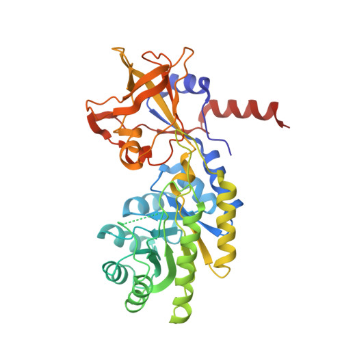
    Lysine decarboxylase (LDC) is a crucial enzyme for acid stress resistance and is also utilized for the biosynthesis of cadaverine, a promising building block for bio-based polyamides. We determined the crystal structure of LDC from Selenomonas ruminantium (SrLDC). SrLDC functions as a dimer and each monomer consists of two distinct domains; a PLP-binding barrel domain and a sheet domain. We also determined the structure of SrLDC in complex with PLP and cadaverine and elucidated the binding mode of cofactor and substrate. Interestingly, compared with the apo-form of SrLDC, the SrLDC in complex with PLP and cadaverine showed a remarkable structural change at the PLP binding site. The PLP binding site of SrLDC contains the highly flexible loops with high b-factors and showed an open-closed conformational change upon the binding of PLP. In fact, SrLDC showed no LDC activity without PLP supplement, and we suggest that highly flexible PLP binding site results in low PLP affinity of SrLDC. In addition, other structurally homologous enzymes also contain the flexible PLP binding site, which indicates that high flexibility at the PLP binding site and low PLP affinity seems to be a common feature of these enzyme family.


  • Organizational Affiliation
    • School of Life Sciences, KNU Creative BioResearch Group, Kyungpook National University, Daegu, 702-701, Republic of Korea.

Macromolecule Content 

  • Total Structure Weight: 44.02 kDa 
  • Atom Count: 3,034 
  • Modeled Residue Count: 381 
  • Deposited Residue Count: 393 
  • Unique protein chains: 1

Macromolecules

Find similar proteins by:|  3D Structure
Entity ID: 1
MoleculeChains  Sequence LengthOrganismDetailsImage
Lysine/ornithine decarboxylase393Selenomonas ruminantiumMutation(s): 0 
Gene Names: ldc
EC: 4.1.1.18 (PDB Primary Data), 4.1.1.17 (UniProt)
UniProt
Find proteins for O50657 (Selenomonas ruminantium)
Explore O50657 
Go to UniProtKB:  O50657
Entity Groups
Sequence Clusters30% Identity50% Identity70% Identity90% Identity95% Identity100% Identity
UniProt GroupO50657
Sequence Annotations
Expand
Reference Sequence

Small Molecules

Ligands 6 Unique
IDChains Name / Formula / InChI Key2D Diagram3D Interactions
PLP

Query on PLP


Download:Ideal Coordinates CCD File
C [auth A]PYRIDOXAL-5'-PHOSPHATE
C8 H10 N O6 P
NGVDGCNFYWLIFO-UHFFFAOYSA-N
PGE

Query on PGE


Download:Ideal Coordinates CCD File
F [auth A]TRIETHYLENE GLYCOL
C6 H14 O4
ZIBGPFATKBEMQZ-UHFFFAOYSA-N
N2P

Query on N2P


Download:Ideal Coordinates CCD File
B [auth A]PENTANE-1,5-DIAMINE
C5 H14 N2
VHRGRCVQAFMJIZ-UHFFFAOYSA-N
GOL

Query on GOL


Download:Ideal Coordinates CCD File
D [auth A],
E [auth A]
GLYCEROL
C3 H8 O3
PEDCQBHIVMGVHV-UHFFFAOYSA-N
MG

Query on MG


Download:Ideal Coordinates CCD File
I [auth A]MAGNESIUM ION
Mg
JLVVSXFLKOJNIY-UHFFFAOYSA-N
NA

Query on NA


Download:Ideal Coordinates CCD File
G [auth A],
H [auth A]
SODIUM ION
Na
FKNQFGJONOIPTF-UHFFFAOYSA-N

Experimental Data & Validation

Experimental Data

  • Method: X-RAY DIFFRACTION
  • Resolution: 2.50 Å
  • R-Value Free:  0.279 (Depositor), 0.279 (DCC) 
  • R-Value Work:  0.198 (Depositor), 0.198 (DCC) 
  • R-Value Observed: 0.200 (Depositor) 
Space Group: P 63 2 2
Unit Cell:
Length ( Å )Angle ( ˚ )
a = 111.734α = 90
b = 111.734β = 90
c = 112.976γ = 120
Software Package:
Software NamePurpose
HKL-2000data collection
HKL-2000data scaling
MOLREPphasing
REFMACrefinement
PDB_EXTRACTdata extraction
HKL-2000data reduction

Structure Validation

View Full Validation Report


Ligand Structure Quality Assessment 


Entry History 

Deposition Data

Revision History  (Full details and data files)

  • Version 1.0: 2016-11-23
    Type: Initial release
  • Version 1.1: 2017-07-19
    Changes: Database references, Derived calculations
  • Version 1.2: 2020-09-09
    Changes: Structure summary
  • Version 1.3: 2023-11-08
    Changes: Data collection, Database references, Refinement description