5GHU | pdb_00005ghu

Crystal structure of YadV chaperone at 1.63 Angstrom


Experimental Data Snapshot

  • Method: X-RAY DIFFRACTION
  • Resolution: 1.63 Å
  • R-Value Free: 
    0.186 (Depositor), 0.190 (DCC) 
  • R-Value Work: 
    0.168 (Depositor), 0.173 (DCC) 
  • R-Value Observed: 
    0.169 (Depositor) 

Starting Models: experimental
View more details

wwPDB Validation 3D Report Full Report

Validation slider image for 5GHU

This is version 1.2 of the entry. See complete history

Literature

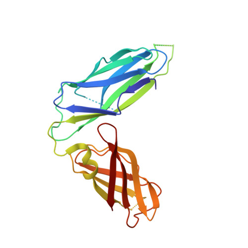
Crystal structure of the usher chaperone YadV reveals a monomer with the proline lock in closed conformation suggestive of an intermediate state.

Pandey, N.K.Verma, G.Kushwaha, G.S.Suar, M.Bhavesh, N.S.

(2020) FEBS Lett 

  • DOI: https://doi.org/10.1002/1873-3468.13883
  • Primary Citation Related Structures: 
    5GHU

  • PubMed Abstract: 

    Cell surface pili assembled by the chaperone-usher (CU) pathway play a crucial role in the adhesion of uropathogenic Escherichia coli. YadV is the chaperone component of the CU pathway of Yad pili. Here, we report the crystal structure of YadV from E. coli. In contrast to major usher chaperones, YadV is a monomer in solution as well as in the crystallographic symmetry, and the monomeric form is a preferred state for interacting with pilus subunits. Moreover, we observed a closed conformation for the proline lock, a crucial structural element for chaperone-pilus subunit interaction. MD simulation shows that the closed state of the proline lock is not energetically stable. Thus, the structure of monomeric YadV with its closed proline lock may serve as an intermediate state to provide suitable access to pilus subunits.


  • Organizational Affiliation
    • Transcription Regulation Group, International Centre for Genetic Engineering and Biotechnology (ICGEB), New Delhi, India.

Macromolecule Content 

  • Total Structure Weight: 24.84 kDa 
  • Atom Count: 1,840 
  • Modeled Residue Count: 201 
  • Deposited Residue Count: 222 
  • Unique protein chains: 1

Macromolecules

Find similar proteins by:|  3D Structure
Entity ID: 1
MoleculeChains  Sequence LengthOrganismDetailsImage
Probable fimbrial chaperone YadV222Escherichia coli K-12Mutation(s): 0 
Gene Names: ecpD
UniProt
Find proteins for P33128 (Escherichia coli (strain K12))
Explore P33128 
Go to UniProtKB:  P33128
Entity Groups
Sequence Clusters30% Identity50% Identity70% Identity90% Identity95% Identity100% Identity
UniProt GroupP33128
Sequence Annotations
Expand
Reference Sequence

Experimental Data & Validation

Experimental Data

  • Method: X-RAY DIFFRACTION
  • Resolution: 1.63 Å
  • R-Value Free:  0.186 (Depositor), 0.190 (DCC) 
  • R-Value Work:  0.168 (Depositor), 0.173 (DCC) 
  • R-Value Observed: 0.169 (Depositor) 
Space Group: C 2 2 21
Unit Cell:
Length ( Å )Angle ( ˚ )
a = 100.28α = 90
b = 127.56β = 90
c = 45.84γ = 90
Software Package:
Software NamePurpose
PHENIXrefinement
MOSFLMdata collection
SCALAdata scaling
PHASERphasing
PDB_EXTRACTdata extraction
iMOSFLMdata processing

Structure Validation

View Full Validation Report


Entry History 

& Funding Information

Deposition Data


Funding OrganizationLocationGrant Number
Department of BiotechnologyIndiaBT/PR6400/BRB/10/113

Revision History  (Full details and data files)

  • Version 1.0: 2017-06-28
    Type: Initial release
  • Version 1.1: 2020-07-22
    Changes: Database references, Structure summary
  • Version 1.2: 2023-11-08
    Changes: Data collection, Database references, Refinement description