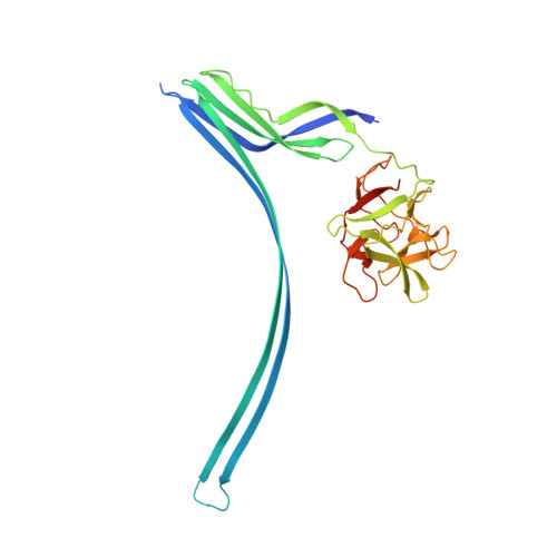
5GAQ

Cryo-EM structure of the Lysenin Pore


Experimental Data Snapshot

  • Method: ELECTRON MICROSCOPY
  • Resolution: 3.10 Å
  • Aggregation State: PARTICLE 
  • Reconstruction Method: SINGLE PARTICLE 

Starting Model: experimental
View more details

wwPDB Validation   3D Report Full Report


This is version 1.8 of the entry. See complete history


Literature

Cryo-EM structure of lysenin pore elucidates membrane insertion by an aerolysin family protein

Bokori-Brown, M.Martin, T.G.Naylor, C.E.Basak, A.K.Titball, R.W.Savva, C.G.

(2016) Nat Commun 7: 11293-11293

  • DOI: https://doi.org/10.1038/ncomms11293
  • Primary Citation of Related Structures:  
    5GAQ

  • PubMed Abstract: 

    Lysenin from the coelomic fluid of the earthworm Eisenia fetida belongs to the aerolysin family of small β-pore-forming toxins (β-PFTs), some members of which are pathogenic to humans and animals. Despite efforts, a high-resolution structure of a channel for this family of proteins has been elusive and therefore the mechanism of activation and membrane insertion remains unclear. Here we determine the pore structure of lysenin by single particle cryo-EM, to 3.1 Å resolution. The nonameric assembly reveals a long β-barrel channel spanning the length of the complex that, unexpectedly, includes the two pre-insertion strands flanking the hypothetical membrane-insertion loop. Examination of other members of the aerolysin family reveals high structural preservation in this region, indicating that the membrane-insertion pathway in this family is conserved. For some toxins, proteolytic activation and pro-peptide removal will facilitate unfolding of the pre-insertion strands, allowing them to form the β-barrel of the channel.


  • Organizational Affiliation

    Biosciences, College of Life and Environmental Sciences, University of Exeter, Stocker Road, Exeter EX4 4QD, UK.


Macromolecules
Find similar proteins by:  (by identity cutoff)  |  3D Structure
Entity ID: 1
MoleculeChains Sequence LengthOrganismDetailsImage
Lysenin
A, B, C, D, E
A, B, C, D, E, F, G, H, I
310Eisenia fetidaMutation(s): 0 
Membrane Entity: Yes 
UniProt
Find proteins for O18423 (Eisenia fetida)
Explore O18423 
Go to UniProtKB:  O18423
Entity Groups  
Sequence Clusters30% Identity50% Identity70% Identity90% Identity95% Identity100% Identity
UniProt GroupO18423
Sequence Annotations
Expand
  • Reference Sequence
Experimental Data & Validation

Experimental Data

  • Method: ELECTRON MICROSCOPY
  • Resolution: 3.10 Å
  • Aggregation State: PARTICLE 
  • Reconstruction Method: SINGLE PARTICLE 
EM Software:
TaskSoftware PackageVersion
RECONSTRUCTIONRELION1.4
MODEL REFINEMENTREFMAC5

Structure Validation

View Full Validation Report


Entry History & Funding Information

Deposition Data


Funding OrganizationLocationGrant Number
Medical Research Council (United Kingdom)United KingdomMC-A021-53019
Wellcome TrustUnited KingdomWT089618MA

Revision History  (Full details and data files)

  • Version 1.0: 2016-04-06
    Type: Initial release
  • Version 1.1: 2016-05-18
    Changes: Database references
  • Version 1.2: 2016-05-25
    Changes: Database references
  • Version 1.3: 2017-06-28
    Changes: Database references
  • Version 1.4: 2017-07-05
    Changes: Derived calculations
  • Version 1.5: 2017-07-19
    Changes: Derived calculations
  • Version 1.6: 2017-08-30
    Changes: Author supporting evidence, Data collection
  • Version 1.7: 2019-10-23
    Changes: Data collection, Other
  • Version 1.8: 2024-05-15
    Changes: Data collection, Database references, Refinement description