5G6U | pdb_00005g6u

Crystal structure of langerin carbohydrate recognition domain with GlcNS6S


Experimental Data Snapshot

  • Method: X-RAY DIFFRACTION
  • Resolution: 1.84 Å
  • R-Value Free: 
    0.180 (Depositor), 0.185 (DCC) 
  • R-Value Work: 
    0.152 (Depositor), 0.157 (DCC) 
  • R-Value Observed: 
    0.153 (Depositor) 

Starting Model: experimental
View more details

wwPDB Validation 3D Report Full Report

Validation slider image for 5G6U

Ligand Structure Quality Assessment 


This is version 2.2 of the entry. See complete history

Literature

Rational-Differential Design of Highly Specific Glycomimetic Ligands: Targeting DC-SIGN and Excluding Langerin Recognition.

Porkolab, V.Chabrol, E.Varga, N.Ordanini, S.Sutkeviciu Te, I.Thepaut, M.Garcia-Jimenez, M.J.Girard, E.Nieto, P.M.Bernardi, A.Fieschi, F.

(2018) ACS Chem Biol 13: 600-608

  • DOI: https://doi.org/10.1021/acschembio.7b00958
  • Primary Citation Related Structures: 
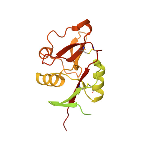
    5G6U

  • PubMed Abstract: 

    At the surface of dendritic cells, C-type lectin receptors (CLRs) allow the recognition of carbohydrate-based PAMPS or DAMPS (pathogen- or danger-associated molecular patterns, respectively) and promote immune response regulation. However, some CLRs are hijacked by viral and bacterial pathogens. Thus, the design of ligands able to target specifically one CLR, to either modulate an immune response or to inhibit a given infection mechanism, has great potential value in therapeutic design. A case study is the selective blocking of DC-SIGN, involved notably in HIV trans-infection of T lymphocytes, without interfering with langerin-mediated HIV clearance. This is a challenging task due to their overlapping carbohydrate specificity. Toward the rational design of DC-SIGN selective ligands, we performed a comparative affinity study between DC-SIGN and langerin with natural ligands. We found that GlcNAc is recognized by both CLRs; however, selective sulfation are shown to increase the selectivity in favor of langerin. With the combination of site-directed mutagenesis and X-ray structural analysis of the langerin/GlcNS6S complex, we highlighted that 6-sulfation of the carbohydrate ligand induced langerin specificity. Additionally, the K313 residue from langerin was identified as a critical feature of its binding site. Using a rational and a differential approach in the study of CLR binding sites, we designed, synthesized, and characterized a new glycomimetic, which is highly specific for DC-SIGN vs langerin. STD NMR, SPR, and ITC characterizations show that compound 7 conserved the overall binding mode of the natural disaccharide while possessing an improved affinity and a strict specificity for DC-SIGN.


  • Organizational Affiliation
    • Univ. Grenoble Alpes , CNRS, CEA, Institut de Biologie Structurale , F-38044 Grenoble , France.

Macromolecule Content 

  • Total Structure Weight: 121.33 kDa 
  • Atom Count: 4,932 
  • Modeled Residue Count: 514 
  • Deposited Residue Count: 1,044 
  • Unique protein chains: 1

Macromolecules

Find similar proteins by:|  3D Structure
Entity ID: 1
MoleculeChains  Sequence LengthOrganismDetailsImage
LANGERIN
A, B, C, D
261Homo sapiensMutation(s): 0 
UniProt & NIH Common Fund Data Resources
Find proteins for Q9UJ71 (Homo sapiens)
Explore Q9UJ71 
Go to UniProtKB:  Q9UJ71
PHAROS:  Q9UJ71
GTEx:  ENSG00000116031 
Entity Groups
Sequence Clusters30% Identity50% Identity70% Identity90% Identity95% Identity100% Identity
UniProt GroupQ9UJ71
Sequence Annotations
Expand
Reference Sequence

Small Molecules

Ligands 5 Unique
IDChains Name / Formula / InChI Key2D Diagram3D Interactions
SGN

Query on SGN


Download:Ideal Coordinates CCD File
CA [auth D],
F [auth A],
L [auth B],
U [auth C]
2-deoxy-6-O-sulfo-2-(sulfoamino)-alpha-D-glucopyranose
C6 H13 N O11 S2
DQTRACMFIGDHSN-UKFBFLRUSA-N
TRP

Query on TRP


Download:Ideal Coordinates CCD File
BA [auth D],
E [auth A],
K [auth B]
TRYPTOPHAN
C11 H12 N2 O2
QIVBCDIJIAJPQS-VIFPVBQESA-N
EU

Query on EU


Download:Ideal Coordinates CCD File
G [auth A]
J [auth A]
M [auth B]
T [auth B]
W [auth C]
G [auth A],
J [auth A],
M [auth B],
T [auth B],
W [auth C],
X [auth C]
EUROPIUM ION
Eu
MGVUQZZTJGLWJV-UHFFFAOYSA-N
CA

Query on CA


Download:Ideal Coordinates CCD File
DA [auth D],
H [auth A],
N [auth B],
V [auth C]
CALCIUM ION
Ca
BHPQYMZQTOCNFJ-UHFFFAOYSA-N
CL

Query on CL


Download:Ideal Coordinates CCD File
AA [auth C]
EA [auth D]
FA [auth D]
GA [auth D]
I [auth A]
AA [auth C],
EA [auth D],
FA [auth D],
GA [auth D],
I [auth A],
O [auth B],
P [auth B],
Q [auth B],
R [auth B],
S [auth B],
Y [auth C],
Z [auth C]
CHLORIDE ION
Cl
VEXZGXHMUGYJMC-UHFFFAOYSA-M

Experimental Data & Validation

Experimental Data

  • Method: X-RAY DIFFRACTION
  • Resolution: 1.84 Å
  • R-Value Free:  0.180 (Depositor), 0.185 (DCC) 
  • R-Value Work:  0.152 (Depositor), 0.157 (DCC) 
  • R-Value Observed: 0.153 (Depositor) 
Space Group: P 42
Unit Cell:
Length ( Å )Angle ( ˚ )
a = 79.483α = 90
b = 79.483β = 90
c = 90.71γ = 90
Software Package:
Software NamePurpose
PHENIXrefinement
XDSdata reduction
SCALAdata scaling
PHASERphasing

Structure Validation

View Full Validation Report


Ligand Structure Quality Assessment 


Entry History 

Deposition Data

Revision History  (Full details and data files)

  • Version 1.0: 2018-02-21
    Type: Initial release
  • Version 1.1: 2018-02-28
    Changes: Derived calculations
  • Version 1.2: 2018-03-28
    Changes: Database references
  • Version 2.0: 2020-07-29
    Type: Remediation
    Reason: Carbohydrate remediation
    Changes: Advisory, Atomic model, Data collection, Derived calculations, Non-polymer description, Structure summary
  • Version 2.1: 2024-01-10
    Changes: Data collection, Database references, Refinement description
  • Version 2.2: 2024-11-13
    Changes: Structure summary