5G5F | pdb_00005g5f

Crystallographic structure of the Tau class glutathione S-transferase MiGSTU in complex with reduced glutathione.


Experimental Data Snapshot

  • Method: X-RAY DIFFRACTION
  • Resolution: 2.30 Å
  • R-Value Free: 
    0.243 (Depositor), 0.242 (DCC) 
  • R-Value Work: 
    0.193 (Depositor), 0.194 (DCC) 
  • R-Value Observed: 
    0.198 (Depositor) 

Starting Model: experimental
View more details

wwPDB Validation 3D Report Full Report

Validation slider image for 5G5F

Ligand Structure Quality Assessment 


This is version 1.3 of the entry. See complete history

Literature

Insights Into Ligand Binding to a Glutathione S-Transferase from Mango: Structure, Thermodynamics and Kinetics

Valenzuela-Chavira, I.Contreras-Vergara, C.A.Arvizu-Flores, A.A.Serrano-Posada, H.Lopez-Zavala, A.A.Garcia-Orozco, K.D.Hernandez-Paredes, J.Rudino-Pinera, E.Stojanoff, V.Sotelo-Mundo, R.R.Islas-Osuna, M.A.

(2017) Biochimie 135: 35

  • DOI: https://doi.org/10.1016/j.biochi.2017.01.005
  • Primary Citation Related Structures: 
    5G5E, 5G5F, 5KEJ

  • PubMed Abstract: 

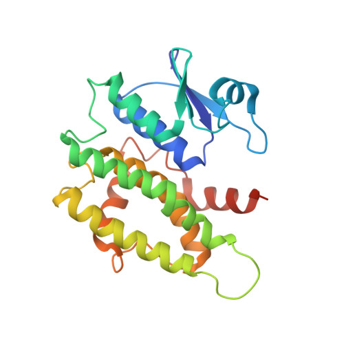
    We studied a mango glutathione S-transferase (GST) (Mangifera indica) bound to glutathione (GSH) and S-hexyl glutathione (GSX). This GST Tau class (MiGSTU) had a molecular mass of 25.5 kDa. MiGSTU Michaelis-Menten kinetic constants were determined for their substrates obtaining a K m , V max and k cat for CDNB of 0.792 mM, 80.58 mM min -1 and 68.49 s -1 respectively and 0.693 mM, 105.32 mM min -1 and 89.57 s -1 , for reduced GSH respectively. MiGSTU had a micromolar affinity towards GSH (5.2 μM) or GSX (7.8 μM). The crystal structure of the MiGSTU in apo or bound to GSH or GSX generated a model that explains the thermodynamic signatures of binding and showed the importance of enthalpic-entropic compensation in ligand binding to Tau-class GST enzymes.


  • Organizational Affiliation
    • Laboratorio de Estructura Biomolecular, Centro de Investigación en Alimentación y Desarrollo, A.C. (CIAD), Hermosillo, Sonora 83304, Mexico; Laboratorio de Genética Molecular de Plantas, Centro de Investigación en Alimentación y Desarrollo, A.C. (CIAD), Hermosillo, Sonora 83304, Mexico.

Macromolecule Content 

  • Total Structure Weight: 26.28 kDa 
  • Atom Count: 1,826 
  • Modeled Residue Count: 219 
  • Deposited Residue Count: 229 
  • Unique protein chains: 1

Macromolecules

Find similar proteins by:|  3D Structure
Entity ID: 1
MoleculeChains  Sequence LengthOrganismDetailsImage
TAU CLASS GLUTATHIONE S-TRANSFERASE229Mangifera indicaMutation(s): 0 
EC: 2.5.1.18
UniProt
Find proteins for A0A1P8NWC2 (Mangifera indica)
Explore A0A1P8NWC2 
Go to UniProtKB:  A0A1P8NWC2
Entity Groups
Sequence Clusters30% Identity50% Identity70% Identity90% Identity95% Identity100% Identity
UniProt GroupA0A1P8NWC2
Sequence Annotations
Expand
Reference Sequence

Biologically Interesting Molecules (External Reference) 

1 Unique

Experimental Data & Validation

Experimental Data

  • Method: X-RAY DIFFRACTION
  • Resolution: 2.30 Å
  • R-Value Free:  0.243 (Depositor), 0.242 (DCC) 
  • R-Value Work:  0.193 (Depositor), 0.194 (DCC) 
  • R-Value Observed: 0.198 (Depositor) 
Space Group: C 1 2 1
Unit Cell:
Length ( Å )Angle ( ˚ )
a = 87.483α = 90
b = 48.24β = 104.16
c = 52.684γ = 90
Software Package:
Software NamePurpose
PHENIXrefinement
PROTEUM2data reduction
SADABSdata scaling
PHASERphasing

Structure Validation

View Full Validation Report


Ligand Structure Quality Assessment 


Entry History 

Deposition Data

Revision History  (Full details and data files)

  • Version 1.0: 2017-02-01
    Type: Initial release
  • Version 1.1: 2018-04-04
    Changes: Data collection
  • Version 1.2: 2018-06-06
    Changes: Data collection, Refinement description
  • Version 1.3: 2024-01-10
    Changes: Data collection, Database references, Derived calculations, Other, Refinement description