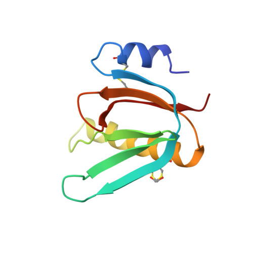
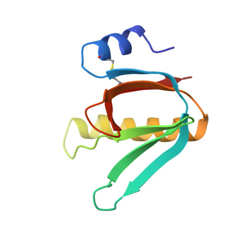
5G3L

ESCHERICHIA COLI HEAT LABILE ENTEROTOXIN TYPE IIB B-PENTAMER COMPLEXED WITH SIALYLATED SUGAR


Experimental Data Snapshot

  • Method: X-RAY DIFFRACTION
  • Resolution: 1.72 Å
  • R-Value Free: 0.197 
  • R-Value Work: 0.171 
  • R-Value Observed: 0.172 

Starting Model: experimental
View more details

wwPDB Validation   3D Report Full Report


Ligand Structure Quality Assessment 


This is version 2.1 of the entry. See complete history


Literature

Biochemical and Structural Characterization of the Novel Sialic Acid-Binding Site of Escherichia Coli Heat-Labile Enterotoxin Lt-Iib.

Zalem, D.Ribeiro, J.P.Varrot, A.Lebens, M.Imberty, A.Teneberg, S.

(2016) Biochem J 473: 3923

  • DOI: https://doi.org/10.1042/BCJ20160575
  • Primary Citation of Related Structures:  
    5G3L

  • PubMed Abstract: 

    The structurally related AB 5 -type heat-labile enterotoxins of Escherichia coli and Vibrio cholerae are classified into two major types. The type I group includes cholera toxin (CT) and E. coli LT-I, whereas the type II subfamily comprises LT-IIa, LT-IIb and LT-IIc. The carbohydrate-binding specificities of LT-IIa, LT-IIb and LT-IIc are distinctive from those of cholera toxin and E. coli LT-I. Whereas CT and LT-I bind primarily to the GM1 ganglioside, LT-IIa binds to gangliosides GD1a, GD1b and GM1, LT-IIb binds to the GD1a and GT1b gangliosides, and LT-IIc binds to GM1, GM2, GM3 and GD1a. These previous studies of the binding properties of type II B-subunits have been focused on ganglio core chain gangliosides. To further define the carbohydrate binding specificity of LT-IIb B-subunits, we have investigated its binding to a collection of gangliosides and non-acid glycosphingolipids with different core chains. A high-affinity binding of LT-IIb B-subunits to gangliosides with a neolacto core chain, such as Neu5Gcα3- and Neu5Acα3-neolactohexaosylceramide, and Neu5Gcα3- and Neu5Acα3-neolactooctaosylceramide was detected. An LT-IIb-binding ganglioside was isolated from human small intestine and characterized as Neu5Acα3-neolactohexaosylceramide. The crystal structure of the B-subunit of LT-IIb with the pentasaccharide moiety of Neu5Acα3-neolactotetraosylceramide (Neu5Ac-nLT: Neu5Acα3Galβ4GlcNAcβ3Galβ4Glc) was determined providing the first information for a sialic-binding site in this subfamily, with clear differences from that of CT and LT-I.


  • Organizational Affiliation

    Institute of Biomedicine, Department of Medical Biochemistry and Cell Biology, The Sahlgrenska Academy, University of Gothenburg, Göteborg, Sweden.


Macromolecules
Find similar proteins by:  (by identity cutoff)  |  3D Structure
Entity ID: 1
MoleculeChains Sequence LengthOrganismDetailsImage
HEAT-LABILE ENTEROTOXIN IIB, B CHAINA [auth D],
B [auth E],
E [auth H]
99Escherichia coli HB101Mutation(s): 0 
UniProt
Find proteins for P43529 (Escherichia coli)
Explore P43529 
Go to UniProtKB:  P43529
Entity Groups  
Sequence Clusters30% Identity50% Identity70% Identity90% Identity95% Identity100% Identity
UniProt GroupP43529
Sequence Annotations
Expand
  • Reference Sequence
Find similar proteins by:  (by identity cutoff)  |  3D Structure
Entity ID: 2
MoleculeChains Sequence LengthOrganismDetailsImage
HEAT-LABILE ENTEROTOXIN IIB, B CHAINC [auth F],
D [auth G]
99Escherichia coli HB101Mutation(s): 0 
UniProt
Find proteins for P43529 (Escherichia coli)
Explore P43529 
Go to UniProtKB:  P43529
Entity Groups  
Sequence Clusters30% Identity50% Identity70% Identity90% Identity95% Identity100% Identity
UniProt GroupP43529
Sequence Annotations
Expand
  • Reference Sequence
Oligosaccharides

Help

Entity ID: 3
MoleculeChains Length2D Diagram Glycosylation3D Interactions
N-acetyl-alpha-neuraminic acid-(2-3)-beta-D-galactopyranoseF [auth A],
G [auth B],
H [auth C]
2N/A
Glycosylation Resources
GlyTouCan:  G30207PZ
GlyCosmos:  G30207PZ
GlyGen:  G30207PZ
Experimental Data & Validation

Experimental Data

  • Method: X-RAY DIFFRACTION
  • Resolution: 1.72 Å
  • R-Value Free: 0.197 
  • R-Value Work: 0.171 
  • R-Value Observed: 0.172 
  • Space Group: P 21 21 21
Unit Cell:
Length ( Å )Angle ( ˚ )
a = 48.564α = 90
b = 68.015β = 90
c = 154.513γ = 90
Software Package:
Software NamePurpose
REFMACrefinement
XDSdata reduction
Aimlessdata scaling
PHASERphasing

Structure Validation

View Full Validation Report


Ligand Structure Quality Assessment 


Entry History 

Deposition Data

Revision History  (Full details and data files)

  • Version 1.0: 2016-09-14
    Type: Initial release
  • Version 1.1: 2016-11-09
    Changes: Database references
  • Version 2.0: 2020-07-29
    Type: Remediation
    Reason: Carbohydrate remediation
    Changes: Advisory, Atomic model, Data collection, Derived calculations, Other, Structure summary
  • Version 2.1: 2024-01-10
    Changes: Data collection, Database references, Derived calculations, Refinement description, Structure summary