5G1X | pdb_00005g1x

Crystal structure of Aurora-A kinase in complex with N-Myc


Experimental Data Snapshot

  • Method: X-RAY DIFFRACTION
  • Resolution: 1.72 Å
  • R-Value Free: 
    0.195 (Depositor), 0.198 (DCC) 
  • R-Value Work: 
    0.176 (Depositor), 0.179 (DCC) 
  • R-Value Observed: 
    0.177 (Depositor) 

Starting Model: experimental
View more details

wwPDB Validation 3D Report Full Report

Validation slider image for 5G1X

Ligand Structure Quality Assessment 


This is version 1.4 of the entry. See complete history

Literature

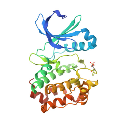
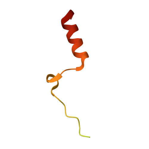
Structural Basis of N-Myc Binding by Aurora-A and its Destabilization by Kinase Inhibitors

Richards, M.Burgess, S.Poon, E.Carstensen, A.Eilers, M.Chesler, L.Bayliss, R.

(2016) Proc Natl Acad Sci U S A 113: 13726

  • DOI: https://doi.org/10.1073/pnas.1610626113
  • Primary Citation Related Structures: 
    5G1X

  • PubMed Abstract: 

    Myc family proteins promote cancer by inducing widespread changes in gene expression. Their rapid turnover by the ubiquitin-proteasome pathway is regulated through phosphorylation of Myc Box I and ubiquitination by the E3 ubiquitin ligase SCF FbxW7 However, N-Myc protein (the product of the MYCN oncogene) is stabilized in neuroblastoma by the protein kinase Aurora-A in a manner that is sensitive to certain Aurora-A-selective inhibitors. Here we identify a direct interaction between the catalytic domain of Aurora-A and a site flanking Myc Box I that also binds SCF FbxW7 We determined the crystal structure of the complex between Aurora-A and this region of N-Myc to 1.72-Å resolution. The structure indicates that the conformation of Aurora-A induced by compounds such as alisertib and CD532 is not compatible with the binding of N-Myc, explaining the activity of these compounds in neuroblastoma cells and providing a rational basis for the design of cancer therapeutics optimized for destabilization of the complex. We also propose a model for the stabilization mechanism in which binding to Aurora-A alters how N-Myc interacts with SCF FbxW7 to disfavor the generation of Lys48-linked polyubiquitin chains.


  • Organizational Affiliation
    • Astbury Centre for Structural and Molecular Biology, Faculty of Biological Sciences, University of Leeds, Leeds LS2 9JT, United Kingdom.

Macromolecule Content 

  • Total Structure Weight: 40.62 kDa 
  • Atom Count: 2,588 
  • Modeled Residue Count: 293 
  • Deposited Residue Count: 348 
  • Unique protein chains: 2

Macromolecules

Find similar proteins by:|  3D Structure
Entity ID: 1
MoleculeChains  Sequence LengthOrganismDetailsImage
AURORA KINASE A285Homo sapiensMutation(s): 2 
EC: 2.7.11.1
UniProt & NIH Common Fund Data Resources
Find proteins for O14965 (Homo sapiens)
Explore O14965 
Go to UniProtKB:  O14965
PHAROS:  O14965
GTEx:  ENSG00000087586 
Entity Groups
Sequence Clusters30% Identity50% Identity70% Identity90% Identity95% Identity100% Identity
UniProt GroupO14965
Sequence Annotations
Expand
Reference Sequence
Find similar proteins by:|  3D Structure
Entity ID: 2
MoleculeChains  Sequence LengthOrganismDetailsImage
N-MYC PROTO-ONCOGENE PROTEIN63Homo sapiensMutation(s): 0 
UniProt & NIH Common Fund Data Resources
Find proteins for P04198 (Homo sapiens)
Explore P04198 
Go to UniProtKB:  P04198
PHAROS:  P04198
GTEx:  ENSG00000134323 
Entity Groups
Sequence Clusters30% Identity50% Identity70% Identity90% Identity95% Identity100% Identity
UniProt GroupP04198
Sequence Annotations
Expand
Reference Sequence

Small Molecules

Experimental Data & Validation

Experimental Data

  • Method: X-RAY DIFFRACTION
  • Resolution: 1.72 Å
  • R-Value Free:  0.195 (Depositor), 0.198 (DCC) 
  • R-Value Work:  0.176 (Depositor), 0.179 (DCC) 
  • R-Value Observed: 0.177 (Depositor) 
Space Group: P 32 2 1
Unit Cell:
Length ( Å )Angle ( ˚ )
a = 86.52α = 90
b = 86.52β = 90
c = 92.21γ = 120
Software Package:
Software NamePurpose
PHENIXrefinement
xia2data reduction
xia2data scaling
PHENIXphasing

Structure Validation

View Full Validation Report


Ligand Structure Quality Assessment 


Entry History 

Deposition Data

Revision History  (Full details and data files)

  • Version 1.0: 2016-11-16
    Type: Initial release
  • Version 1.1: 2016-11-23
    Changes: Structure summary
  • Version 1.2: 2016-12-14
    Changes: Database references
  • Version 1.3: 2024-01-10
    Changes: Data collection, Database references, Derived calculations, Other, Refinement description
  • Version 1.4: 2024-10-16
    Changes: Structure summary