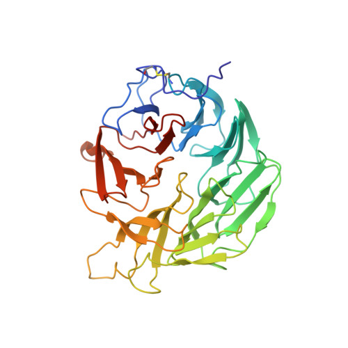
5FZP

Structure of the dispase autolysis inducing protein from Streptomyces mobaraensis


Experimental Data Snapshot

  • Method: X-RAY DIFFRACTION
  • Resolution: 1.70 Å
  • R-Value Free: 0.191 
  • R-Value Work: 0.159 
  • R-Value Observed: 0.161 

wwPDB Validation   3D Report Full Report


This is version 1.3 of the entry. See complete history


Literature

Structure of the Dispase Autolysis Inducing Protein from Streptomyces Mobaraensis and Glutamine Cross-Linking Sites for Transglutaminase

Fiebig, D.Schmelz, S.Zindel, S.Ehret, V.Beck, J.Ebenig, A.Ehret, M.Froels, S.Pfeifer, F.Kolmar, H.Fuchsbauer, H.L.Scrima, A.

(2016) J Biol Chem 291: 20417

  • DOI: https://doi.org/10.1074/jbc.M116.731109
  • Primary Citation of Related Structures:  
    5FZP

  • PubMed Abstract: 

    Transglutaminase from Streptomyces mobaraensis (MTG) is an important enzyme for cross-linking and modifying proteins. An intrinsic substrate of MTG is the dispase autolysis-inducing protein (DAIP). The amino acid sequence of DAIP contains 5 potential glutamines and 10 lysines for MTG-mediated cross-linking. The aim of the study was to determine the structure and glutamine cross-linking sites of the first physiological MTG substrate. A production procedure was established in Escherichia coli BL21 (DE3) to obtain high yields of recombinant DAIP. DAIP variants were prepared by replacing four of five glutamines for asparagines in various combinations via site-directed mutagenesis. Incorporation of biotin cadaverine revealed a preference of MTG for the DAIP glutamines in the order of Gln-39 ≫ Gln-298 > Gln-345 ∼ Gln-65 ≫ Gln-144. In the structure of DAIP the preferred glutamines do cluster at the top of the seven-bladed β-propeller. This suggests a targeted cross-linking of DAIP by MTG that may occur after self-assembly in the bacterial cell wall. Based on our biochemical and structural data of the first physiological MTG substrate, we further provide novel insight into determinants of MTG-mediated modification, specificity, and efficiency.


  • Organizational Affiliation

    Department of Chemical Engineering and Biotechnology, University of Applied Sciences of Darmstadt, 64287 Darmstadt, Germany, and the Department of Chemistry and.


Macromolecules
Find similar proteins by:  (by identity cutoff)  |  3D Structure
Entity ID: 1
MoleculeChains Sequence LengthOrganismDetailsImage
DISPASE AUTOLYSIS-INDUCING PROTEIN
A, B
348Streptomyces mobaraensisMutation(s): 0 
UniProt
Find proteins for P84908 (Streptomyces mobaraensis)
Explore P84908 
Go to UniProtKB:  P84908
Entity Groups  
Sequence Clusters30% Identity50% Identity70% Identity90% Identity95% Identity100% Identity
UniProt GroupP84908
Sequence Annotations
Expand
  • Reference Sequence
Small Molecules
Ligands 2 Unique
IDChains Name / Formula / InChI Key2D Diagram3D Interactions
GOL
Query on GOL

Download Ideal Coordinates CCD File 
C [auth A]
D [auth A]
E [auth A]
F [auth A]
I [auth B]
C [auth A],
D [auth A],
E [auth A],
F [auth A],
I [auth B],
J [auth B],
K [auth B]
GLYCEROL
C3 H8 O3
PEDCQBHIVMGVHV-UHFFFAOYSA-N
CA
Query on CA

Download Ideal Coordinates CCD File 
G [auth A],
H [auth A],
L [auth B],
M [auth B]
CALCIUM ION
Ca
BHPQYMZQTOCNFJ-UHFFFAOYSA-N
Experimental Data & Validation

Experimental Data

  • Method: X-RAY DIFFRACTION
  • Resolution: 1.70 Å
  • R-Value Free: 0.191 
  • R-Value Work: 0.159 
  • R-Value Observed: 0.161 
  • Space Group: P 21 21 21
Unit Cell:
Length ( Å )Angle ( ˚ )
a = 61.01α = 90
b = 101.13β = 90
c = 114.66γ = 90
Software Package:
Software NamePurpose
PHENIXrefinement
XDSdata reduction
XSCALEdata scaling
HKL2MAPphasing

Structure Validation

View Full Validation Report


Entry History 

Deposition Data

Revision History  (Full details and data files)

  • Version 1.0: 2016-08-10
    Type: Initial release
  • Version 1.1: 2016-08-17
    Changes: Database references
  • Version 1.2: 2016-08-24
    Changes: Derived calculations
  • Version 1.3: 2016-10-05
    Changes: Database references