5FW4 | pdb_00005fw4

Structure of Thermobifida fusca DyP-type Peroxidase and Activity towards Kraft Lignin and Lignin Model Compounds


Experimental Data Snapshot

  • Method: X-RAY DIFFRACTION
  • Resolution: 1.80 Å
  • R-Value Free: 
    0.249 (Depositor), 0.251 (DCC) 
  • R-Value Work: 
    0.204 (Depositor), 0.209 (DCC) 
  • R-Value Observed: 
    0.206 (Depositor) 

Starting Model: experimental
View more details

wwPDB Validation 3D Report Full Report

Validation slider image for 5FW4

Ligand Structure Quality Assessment 


This is version 1.1 of the entry. See complete history

Literature

Structure of Thermobifida Fusca Dyp-Type Peroxidase and Activity Towards Kraft Lignin and Lignin Model Compounds.

Rahmanpour, R.Rea, D.Jamshidi, S.Fulop, V.Bugg, T.D.H.

(2016) Arch Biochem Biophys 594: 54

  • DOI: https://doi.org/10.1016/j.abb.2016.02.019
  • Primary Citation Related Structures: 
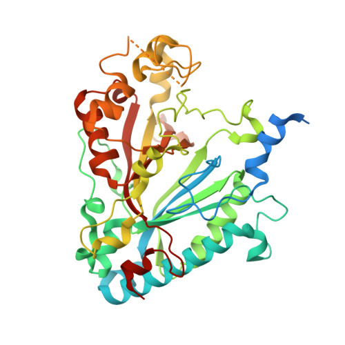
    5FW4

  • PubMed Abstract: 

    A Dyp-type peroxidase enzyme from thermophilic cellulose degrader Thermobifida fusca (TfuDyP) was investigated for catalytic ability towards lignin oxidation. TfuDyP was characterised kinetically against a range of phenolic substrates, and a compound I reaction intermediate was observed via pre-steady state kinetic analysis at λmax 404 nm. TfuDyP showed reactivity towards Kraft lignin, and was found to oxidise a β-aryl ether lignin model compound, forming an oxidised dimer. A crystal structure of TfuDyP was determined, to 1.8 Å resolution, which was found to contain a diatomic oxygen ligand bound to the heme centre, positioned close to active site residues Asp-203 and Arg-315. The structure contains two channels providing access to the heme cofactor for organic substrates and hydrogen peroxide. Site-directed mutant D203A showed no activity towards phenolic substrates, but reduced activity towards ABTS, while mutant R315Q showed no activity towards phenolic substrates, nor ABTS.


  • Organizational Affiliation
    • Department of Chemistry, University of Warwick, Gibbet Hill Road, Coventry CV4 7AL, UK.

Macromolecule Content 

  • Total Structure Weight: 93.26 kDa 
  • Atom Count: 6,088 
  • Modeled Residue Count: 736 
  • Deposited Residue Count: 860 
  • Unique protein chains: 1

Macromolecules

Find similar proteins by:|  3D Structure
Entity ID: 1
MoleculeChains  Sequence LengthOrganismDetailsImage
DYE-DECOLORIZING PEROXIDASE TFU_3078
A, B
430Thermobifida fusca YXMutation(s): 0 
EC: 1.11.1.19
UniProt
Find proteins for Q47KB1 (Thermobifida fusca (strain YX))
Explore Q47KB1 
Go to UniProtKB:  Q47KB1
Entity Groups
Sequence Clusters30% Identity50% Identity70% Identity90% Identity95% Identity100% Identity
UniProt GroupQ47KB1
Sequence Annotations
Expand
Reference Sequence

Experimental Data & Validation

Experimental Data

  • Method: X-RAY DIFFRACTION
  • Resolution: 1.80 Å
  • R-Value Free:  0.249 (Depositor), 0.251 (DCC) 
  • R-Value Work:  0.204 (Depositor), 0.209 (DCC) 
  • R-Value Observed: 0.206 (Depositor) 
Space Group: P 21 21 2
Unit Cell:
Length ( Å )Angle ( ˚ )
a = 143.36α = 90
b = 110.21β = 90
c = 46.73γ = 90
Software Package:
Software NamePurpose
REFMACrefinement
XDSdata reduction
XDSdata scaling
PHASERphasing

Structure Validation

View Full Validation Report


Ligand Structure Quality Assessment 


Entry History 

Deposition Data

Revision History  (Full details and data files)

  • Version 1.0: 2016-04-06
    Type: Initial release
  • Version 1.1: 2024-01-10
    Changes: Data collection, Database references, Derived calculations, Other, Refinement description