5FV1 | pdb_00005fv1

Crystal structure of hVEGF in complex with VK domain antibody


Experimental Data Snapshot

  • Method: X-RAY DIFFRACTION
  • Resolution: 2.70 Å
  • R-Value Free: 
    0.261 (Depositor), 0.259 (DCC) 
  • R-Value Work: 
    0.205 (Depositor), 0.207 (DCC) 
  • R-Value Observed: 
    0.208 (Depositor) 

wwPDB Validation 3D Report Full Report

Validation slider image for 5FV1

This is version 1.4 of the entry. See complete history

Literature

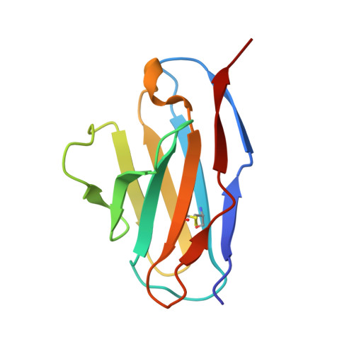
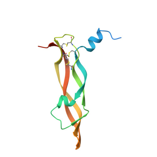
Novel Interaction Mechanism of a Domain Antibody Based Inhibitor of Human Vascular Endothelial Growth Factor with Greater Potency Than Ranibizumab and Bevacizumab and Improved Capacity Over Aflibercept.

Walker, A.Chung, C.Neu, M.Burman, M.Batuwangala, T.Jones, G.Tang, C.Steward, M.Mullin, M.Tournier, N.Lewis, A.Korczynska, J.Chung, V.Catchpole, I.

(2016) J Biological Chem 291: 5500

  • DOI: https://doi.org/10.1074/jbc.M115.691162
  • Primary Citation Related Structures: 
    5FV1, 5FV2

  • PubMed Abstract: 

    A potent VEGF inhibitor with novel antibody architecture and antigen binding mode has been developed. The molecule, hereafter referred to as VEGF dual dAb (domain antibody), was evaluated in vitro for binding to VEGF and for potency in VEGF-driven models and compared with other anti-VEGF biologics that have been used in ocular anti-angiogenic therapeutic regimes. VEGF dual dAb is more potent than bevacizumab and ranibizumab for VEGF binding, inhibition of VEGF receptor binding assays (RBAs), and VEGF-driven in vitro models of angiogenesis and displays comparable inhibition to aflibercept (Eylea). VEGF dual dAb is dimeric, and each monomer contains two distinct anti-VEGF domain antibodies attached via linkers to a human IgG1 Fc domain. Mechanistically, the enhanced in vitro potency of VEGF dual dAb, in comparison to other anti-VEGF biologics, can be explained by increased binding stoichiometry. A consistent model of the target engagement has been built based on the x-ray complexes of each of the two isolated domain antibodies with the VEGF antigen.


  • Organizational Affiliation
    • From BioPharm Innovation.

Macromolecule Content 

  • Total Structure Weight: 51.18 kDa 
  • Atom Count: 3,329 
  • Modeled Residue Count: 410 
  • Deposited Residue Count: 448 
  • Unique protein chains: 2

Macromolecules

Find similar proteins by:|  3D Structure
Entity ID: 1
MoleculeChains  Sequence LengthOrganismDetailsImage
VK DOMAIN ANTIBODYA [auth L],
B [auth M]
108Homo sapiensMutation(s): 0 
Find similar proteins by:|  3D Structure
Entity ID: 2
MoleculeChains  Sequence LengthOrganismDetailsImage
VASCULAR ENDOTHELIAL GROWTH FACTOR AC [auth V],
D [auth W]
116Homo sapiensMutation(s): 0 
UniProt & NIH Common Fund Data Resources
Find proteins for P15692 (Homo sapiens)
Explore P15692 
Go to UniProtKB:  P15692
PHAROS:  P15692
GTEx:  ENSG00000112715 
Entity Groups
Sequence Clusters30% Identity50% Identity70% Identity90% Identity95% Identity100% Identity
UniProt GroupP15692
Sequence Annotations
Expand
Reference Sequence

Experimental Data & Validation

Experimental Data

  • Method: X-RAY DIFFRACTION
  • Resolution: 2.70 Å
  • R-Value Free:  0.261 (Depositor), 0.259 (DCC) 
  • R-Value Work:  0.205 (Depositor), 0.207 (DCC) 
  • R-Value Observed: 0.208 (Depositor) 
Space Group: I 2 2 2
Unit Cell:
Length ( Å )Angle ( ˚ )
a = 61.221α = 90
b = 147.128β = 90
c = 175.171γ = 90
Software Package:
Software NamePurpose
REFMACrefinement
XDSdata reduction
SCALAdata scaling
PHASERphasing

Structure Validation

View Full Validation Report


Entry History 

Deposition Data

Revision History  (Full details and data files)

  • Version 1.0: 2016-02-17
    Type: Initial release
  • Version 1.1: 2016-03-23
    Changes: Database references
  • Version 1.2: 2018-04-25
    Changes: Data collection
  • Version 1.3: 2019-05-15
    Changes: Data collection, Experimental preparation
  • Version 1.4: 2024-11-13
    Changes: Data collection, Database references, Other, Structure summary