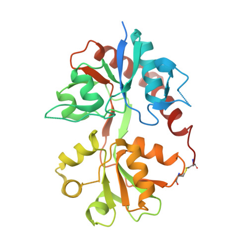
5FTH

Crystal structure of the GluA2 K738M-T744K LBD in complex with glutamate (zinc form)


Experimental Data Snapshot

  • Method: X-RAY DIFFRACTION
  • Resolution: 2.90 Å
  • R-Value Free: 0.283 
  • R-Value Work: 0.243 
  • R-Value Observed: 0.245 

Starting Model: experimental
View more details

wwPDB Validation   3D Report Full Report


This is version 1.4 of the entry. See complete history


Literature

Distinct Structural Pathways Coordinate the Activation of Ampa Receptor-Auxiliary Subunit Complexes.

Dawe, G.B.Musgaard, M.Aurousseau, M.R.P.Nayeem, N.Green, T.Biggin, P.C.Bowie, D.

(2016) Neuron 89: 1264

  • DOI: https://doi.org/10.1016/j.neuron.2016.01.038
  • Primary Citation of Related Structures:  
    5FTH, 5FTI

  • PubMed Abstract: 

    Neurotransmitter-gated ion channels adopt different gating modes to fine-tune signaling at central synapses. At glutamatergic synapses, high and low activity of AMPA receptors (AMPARs) is observed when pore-forming subunits coassemble with or without auxiliary subunits, respectively. Whether a common structural pathway accounts for these different gating modes is unclear. Here, we identify two structural motifs that determine the time course of AMPAR channel activation. A network of electrostatic interactions at the apex of the AMPAR ligand-binding domain (LBD) is essential for gating by pore-forming subunits, whereas a conserved motif on the lower, D2 lobe of the LBD prolongs channel activity when auxiliary subunits are present. Accordingly, channel activity is almost entirely abolished by elimination of the electrostatic network but restored via auxiliary protein interactions at the D2 lobe. In summary, we propose that activation of native AMPAR complexes is coordinated by distinct structural pathways, favored by the association/dissociation of auxiliary subunits.


  • Organizational Affiliation

    Integrated Program in Neuroscience, McGill University, Montréal, QC H3A 2B4, Canada; Department of Pharmacology and Therapeutics, McGill University, Montréal, QC H3G 1Y6, Canada.


Macromolecules
Find similar proteins by:  (by identity cutoff)  |  3D Structure
Entity ID: 1
MoleculeChains Sequence LengthOrganismDetailsImage
GLUTAMATE RECEPTOR 2
A, B, C
291Rattus norvegicusMutation(s): 5 
UniProt
Find proteins for P19491 (Rattus norvegicus)
Explore P19491 
Go to UniProtKB:  P19491
Entity Groups  
Sequence Clusters30% Identity50% Identity70% Identity90% Identity95% Identity100% Identity
UniProt GroupP19491
Sequence Annotations
Expand
  • Reference Sequence
Small Molecules
Ligands 2 Unique
IDChains Name / Formula / InChI Key2D Diagram3D Interactions
GLU
Query on GLU

Download Ideal Coordinates CCD File 
D [auth A],
G [auth B],
J [auth C]
GLUTAMIC ACID
C5 H9 N O4
WHUUTDBJXJRKMK-VKHMYHEASA-N
ZN
Query on ZN

Download Ideal Coordinates CCD File 
E [auth A],
F [auth A],
H [auth B],
I [auth B],
K [auth C]
ZINC ION
Zn
PTFCDOFLOPIGGS-UHFFFAOYSA-N
Experimental Data & Validation

Experimental Data

  • Method: X-RAY DIFFRACTION
  • Resolution: 2.90 Å
  • R-Value Free: 0.283 
  • R-Value Work: 0.243 
  • R-Value Observed: 0.245 
  • Space Group: P 2 21 21
Unit Cell:
Length ( Å )Angle ( ˚ )
a = 46.375α = 90
b = 110.523β = 90
c = 167.245γ = 90
Software Package:
Software NamePurpose
XDSdata reduction
Aimlessdata scaling
REFMACphasing
PHENIXphasing
PHENIXrefinement

Structure Validation

View Full Validation Report


Entry History 

Deposition Data

Revision History  (Full details and data files)

  • Version 1.0: 2016-02-03
    Type: Initial release
  • Version 1.1: 2016-03-16
    Changes: Database references
  • Version 1.2: 2016-03-30
    Changes: Database references
  • Version 1.3: 2024-01-10
    Changes: Data collection, Database references, Derived calculations, Other, Refinement description
  • Version 1.4: 2024-11-13
    Changes: Structure summary