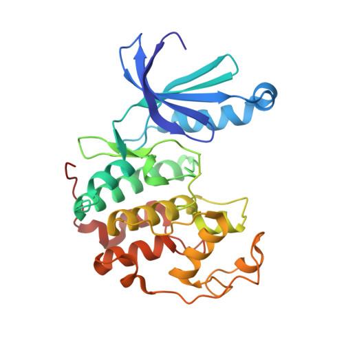
5FP6

Structure of cyclin-dependent kinase 2 with small-molecule ligand 3-(4,7-dichloro-1H-indol-3-yl)prop-2-yn-1-ol (AT17833) in an alternate binding site.


Experimental Data Snapshot

  • Method: X-RAY DIFFRACTION
  • Resolution: 1.85 Å
  • R-Value Free: 0.240 
  • R-Value Work: 0.207 
  • R-Value Observed: 0.209 

Starting Model: experimental
View more details

wwPDB Validation   3D Report Full Report


Ligand Structure Quality Assessment 


This is version 1.3 of the entry. See complete history


Literature

Detection of Secondary Binding Sites in Proteins Using Fragment Screening.

Ludlow, R.F.Verdonk, M.L.Saini, H.K.Tickle, I.J.Jhoti, H.

(2015) Proc Natl Acad Sci U S A 112: 15910

  • DOI: https://doi.org/10.1073/pnas.1518946112
  • Primary Citation of Related Structures:  
    5FP5, 5FP6, 5FPD, 5FPE, 5FPM, 5FPN, 5FPO, 5FPR, 5FPS, 5FPT, 5FPY

  • PubMed Abstract: 

    Proteins need to be tightly regulated as they control biological processes in most normal cellular functions. The precise mechanisms of regulation are rarely completely understood but can involve binding of endogenous ligands and/or partner proteins at specific locations on a protein that can modulate function. Often, these additional secondary binding sites appear separate to the primary binding site, which, for example for an enzyme, may bind a substrate. In previous work, we have uncovered several examples in which secondary binding sites were discovered on proteins using fragment screening approaches. In each case, we were able to establish that the newly identified secondary binding site was biologically relevant as it was able to modulate function by the binding of a small molecule. In this study, we investigate how often secondary binding sites are located on proteins by analyzing 24 protein targets for which we have performed a fragment screen using X-ray crystallography. Our analysis shows that, surprisingly, the majority of proteins contain secondary binding sites based on their ability to bind fragments. Furthermore, sequence analysis of these previously unknown sites indicate high conservation, which suggests that they may have a biological function, perhaps via an allosteric mechanism. Comparing the physicochemical properties of the secondary sites with known primary ligand binding sites also shows broad similarities indicating that many of the secondary sites may be druggable in nature with small molecules that could provide new opportunities to modulate potential therapeutic targets.


  • Organizational Affiliation

    Astex Pharmaceuticals, Cambridge CB4 0QA, United Kingdom.


Macromolecules
Find similar proteins by:  (by identity cutoff)  |  3D Structure
Entity ID: 1
MoleculeChains Sequence LengthOrganismDetailsImage
CYCLIN-DEPENDENT KINASE 2298Homo sapiensMutation(s): 0 
EC: 2.7.11.22
UniProt & NIH Common Fund Data Resources
Find proteins for P24941 (Homo sapiens)
Explore P24941 
Go to UniProtKB:  P24941
PHAROS:  P24941
GTEx:  ENSG00000123374 
Entity Groups  
Sequence Clusters30% Identity50% Identity70% Identity90% Identity95% Identity100% Identity
UniProt GroupP24941
Sequence Annotations
Expand
  • Reference Sequence
Small Molecules
Ligands 1 Unique
IDChains Name / Formula / InChI Key2D Diagram3D Interactions
MFZ
Query on MFZ

Download Ideal Coordinates CCD File 
B [auth A],
C [auth A]
3-(4,7-dichloro-1H-indol-3-yl)prop-2-yn-1-ol
C11 H7 Cl2 N O
VIZAOCOKGQUJRZ-UHFFFAOYSA-N
Experimental Data & Validation

Experimental Data

  • Method: X-RAY DIFFRACTION
  • Resolution: 1.85 Å
  • R-Value Free: 0.240 
  • R-Value Work: 0.207 
  • R-Value Observed: 0.209 
  • Space Group: P 21 21 21
Unit Cell:
Length ( Å )Angle ( ˚ )
a = 53.917α = 90
b = 72.214β = 90
c = 72.198γ = 90
Software Package:
Software NamePurpose
BUSTERrefinement
d*TREKdata reduction
DTSCALEdata scaling
CSEARCHphasing

Structure Validation

View Full Validation Report


Ligand Structure Quality Assessment 


Entry History 

Deposition Data

Revision History  (Full details and data files)

  • Version 1.0: 2015-12-23
    Type: Initial release
  • Version 1.1: 2016-01-13
    Changes: Database references
  • Version 1.2: 2021-04-28
    Changes: Data collection, Derived calculations, Other
  • Version 1.3: 2024-01-10
    Changes: Data collection, Database references, Refinement description