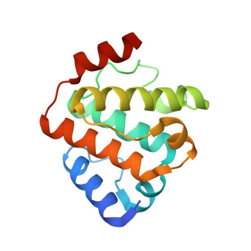
5FMU

MmIFT54 CH-domain


Experimental Data Snapshot

  • Method: X-RAY DIFFRACTION
  • Resolution: 1.59 Å
  • R-Value Free: 0.214 
  • R-Value Work: 0.183 
  • R-Value Observed: 0.184 

Starting Model: experimental
View more details

wwPDB Validation   3D Report Full Report


This is version 1.3 of the entry. See complete history


Literature

Intraflagellar Transport Proteins 172, 80, 57, 54, 38, and 20 Form a Stable Tubulin-Binding Ift-B2 Complex.

Taschner, M.Weber, K.Mourao, A.Vetter, M.Awasthi, M.Stiegler, M.Bhogaraju, S.Lorentzen, E.

(2016) EMBO J 35: 773

  • DOI: https://doi.org/10.15252/embj.201593164
  • Primary Citation of Related Structures:  
    5FMR, 5FMS, 5FMT, 5FMU

  • PubMed Abstract: 

    Intraflagellar transport (IFT) relies on the IFT complex and is required for ciliogenesis. The IFT-B complex consists of 9-10 stably associated core subunits and six "peripheral" subunits that were shown to dissociate from the core structure at moderate salt concentration. We purified the six "peripheral"IFT-B subunits of Chlamydomonas reinhardtiias recombinant proteins and show that they form a stable complex independently of the IFT-B core. We suggest a nomenclature of IFT-B1 (core) and IFT-B2 (peripheral) for the two IFT-B subcomplexes. We demonstrate that IFT88, together with the N-terminal domain of IFT52, is necessary to bridge the interaction between IFT-B1 and B2. The crystal structure of IFT52N reveals highly conserved residues critical for IFT-B1/IFT-B2 complex formation. Furthermore, we show that of the three IFT-B2 subunits containing a calponin homology (CH) domain (IFT38, 54, and 57), only IFT54 binds αβ-tubulin as a potential IFT cargo, whereas the CH domains of IFT38 and IFT57 mediate the interaction with IFT80 and IFT172, respectively. Crystal structures of IFT54 CH domains reveal that tubulin binding is mediated by basic surface-exposed residues.


  • Organizational Affiliation

    Department of Structural Cell Biology, Max-Planck-Institute of Biochemistry, Martinsried, Germany.


Macromolecules
Find similar proteins by:  (by identity cutoff)  |  3D Structure
Entity ID: 1
MoleculeChains Sequence LengthOrganismDetailsImage
TRAF3-INTERACTING PROTEIN 1
A, B, C, D
137Mus musculusMutation(s): 0 
UniProt & NIH Common Fund Data Resources
Find proteins for Q149C2 (Mus musculus)
Explore Q149C2 
Go to UniProtKB:  Q149C2
IMPC:  MGI:1921269
Entity Groups  
Sequence Clusters30% Identity50% Identity70% Identity90% Identity95% Identity100% Identity
UniProt GroupQ149C2
Sequence Annotations
Expand
  • Reference Sequence
Small Molecules
Ligands 1 Unique
IDChains Name / Formula / InChI Key2D Diagram3D Interactions
MES
Query on MES

Download Ideal Coordinates CCD File 
E [auth C]2-(N-MORPHOLINO)-ETHANESULFONIC ACID
C6 H13 N O4 S
SXGZJKUKBWWHRA-UHFFFAOYSA-N
Experimental Data & Validation

Experimental Data

  • Method: X-RAY DIFFRACTION
  • Resolution: 1.59 Å
  • R-Value Free: 0.214 
  • R-Value Work: 0.183 
  • R-Value Observed: 0.184 
  • Space Group: P 1
Unit Cell:
Length ( Å )Angle ( ˚ )
a = 37.93α = 109.17
b = 59.39β = 105.15
c = 61.18γ = 89.65
Software Package:
Software NamePurpose
PHENIXrefinement
PHENIXphasing

Structure Validation

View Full Validation Report


Entry History 

Deposition Data

Revision History  (Full details and data files)

  • Version 1.0: 2016-03-09
    Type: Initial release
  • Version 1.1: 2016-04-13
    Changes: Database references
  • Version 1.2: 2017-08-23
    Changes: Data collection
  • Version 1.3: 2024-01-10
    Changes: Data collection, Database references, Derived calculations, Other, Refinement description