5FLW | pdb_00005flw

Crystal structure of putative exo-beta-1,3-galactanase from Bifidobacterium bifidum s17


Experimental Data Snapshot

  • Method: X-RAY DIFFRACTION
  • Resolution: 1.40 Å
  • R-Value Free: 
    0.183 (Depositor), 0.186 (DCC) 
  • R-Value Work: 
    0.162 (Depositor), 0.163 (DCC) 
  • R-Value Observed: 
    0.162 (Depositor) 

Starting Model: experimental
View more details

wwPDB Validation 3D Report Full Report

Validation slider image for 5FLW

This is version 1.5 of the entry. See complete history

Literature

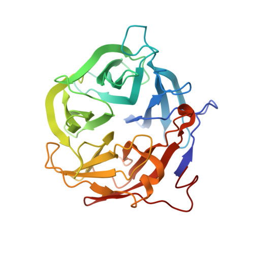
Crystal structure of a putative exo-beta-1,3-galactanase from Bifidobacterium bifidum S17.

Godoy, A.S.de Lima, M.Z.Camilo, C.M.Polikarpov, I.

(2016) Acta Crystallogr F Struct Biol Commun 72: 288-293

  • DOI: https://doi.org/10.1107/S2053230X16003617
  • Primary Citation Related Structures: 
    5FLW

  • PubMed Abstract: 

    Given the current interest in second-generation biofuels, carbohydrate-active enzymes have become the most important tool to overcome the structural recalcitrance of the plant cell wall. While some glycoside hydrolase families have been exhaustively described, others remain poorly characterized, especially with regard to structural information. The family 43 glycoside hydrolases are a diverse group of inverting enzymes; the available structure information on these enzymes is mainly from xylosidases and arabinofuranosidase. Currently, only one structure of an exo-β-1,3-galactanase is available. Here, the production, crystallization and structure determination of a putative exo-β-1,3-galactanase from Bifidobacterium bifidum S17 (BbGal43A) are described. BbGal43A was successfully produced and showed activity towards synthetic galactosides. BbGal43A was subsequently crystallized and data were collected to 1.4 Å resolution. The structure shows a single-domain molecule, differing from known homologues, and crystal contact analysis predicts the formation of a dimer in solution. Further biochemical studies are necessary to elucidate the differences between BbGal43A and its characterized homologues.


  • Organizational Affiliation
    • Departamento de Física em São Carlos, Universidade de São Paulo, Avenida Trabalhador Saocarlense 400, 13560-970 São Carlos-SP, Brazil.

Macromolecule Content 

  • Total Structure Weight: 68.3 kDa 
  • Atom Count: 6,144 
  • Modeled Residue Count: 604 
  • Deposited Residue Count: 608 
  • Unique protein chains: 1

Macromolecules

Find similar proteins by:|  3D Structure
Entity ID: 1
MoleculeChains  Sequence LengthOrganismDetailsImage
EXO-BETA-1,3-GALACTANASE
A, B
304Bifidobacterium bifidum S17Mutation(s): 0 
EC: 3.2.1.145

Small Molecules

Ligands 1 Unique
IDChains Name / Formula / InChI Key2D Diagram3D Interactions
BTB

Query on BTB


Download:Ideal Coordinates CCD File
C [auth B]2-[BIS-(2-HYDROXY-ETHYL)-AMINO]-2-HYDROXYMETHYL-PROPANE-1,3-DIOL
C8 H19 N O5
OWMVSZAMULFTJU-UHFFFAOYSA-N

Experimental Data & Validation

Experimental Data

  • Method: X-RAY DIFFRACTION
  • Resolution: 1.40 Å
  • R-Value Free:  0.183 (Depositor), 0.186 (DCC) 
  • R-Value Work:  0.162 (Depositor), 0.163 (DCC) 
  • R-Value Observed: 0.162 (Depositor) 
Space Group: P 21 21 21
Unit Cell:
Length ( Å )Angle ( ˚ )
a = 64.926α = 90
b = 67.343β = 90
c = 202.126γ = 90
Software Package:
Software NamePurpose
PHENIXrefinement
PHASERphasing

Structure Validation

View Full Validation Report


Entry History 

Deposition Data

Revision History  (Full details and data files)

  • Version 1.0: 2015-12-23
    Type: Initial release
  • Version 1.1: 2016-03-30
    Changes: Database references, Structure summary
  • Version 1.2: 2016-04-13
    Changes: Database references
  • Version 1.3: 2016-04-20
    Changes: Database references
  • Version 1.4: 2017-05-10
    Changes: Database references
  • Version 1.5: 2024-01-10
    Changes: Data collection, Database references, Derived calculations, Other, Refinement description