5FIB | pdb_00005fib

Open form of murine Acid Sphingomyelinase


Experimental Data Snapshot

  • Method: X-RAY DIFFRACTION
  • Resolution: 2.80 Å
  • R-Value Free: 
    0.237 (Depositor), 0.238 (DCC) 
  • R-Value Work: 
    0.190 (Depositor), 0.190 (DCC) 
  • R-Value Observed: 
    0.192 (Depositor) 

Starting Model: experimental
View more details

wwPDB Validation 3D Report Full Report

Validation slider image for 5FIB

Ligand Structure Quality Assessment 


This is version 2.2 of the entry. See complete history

Literature

Crystal structure of mammalian acid sphingomyelinase.

Gorelik, A.Illes, K.Heinz, L.X.Superti-Furga, G.Nagar, B.

(2016) Nat Commun 7: 12196-12196

  • DOI: https://doi.org/10.1038/ncomms12196
  • Primary Citation Related Structures: 
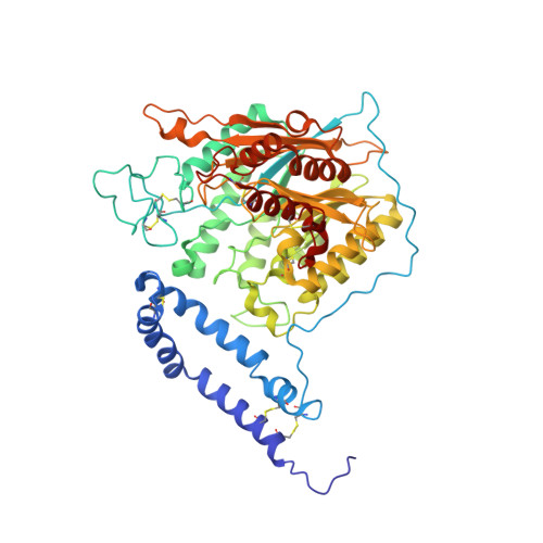
    5FI9, 5FIB, 5FIC, 5HQN

  • PubMed Abstract: 

    Acid sphingomyelinase (ASMase, ASM, SMPD1) converts sphingomyelin into ceramide, modulating membrane properties and signal transduction. Inactivating mutations in ASMase cause Niemann-Pick disease, and its inhibition is also beneficial in models of depression and cancer. To gain a better understanding of this critical therapeutic target, we determined crystal structures of mammalian ASMase in various conformations. The catalytic domain adopts a calcineurin-like fold with two zinc ions and a hydrophobic track leading to the active site. Strikingly, the membrane interacting saposin domain assumes either a closed globular conformation independent from the catalytic domain, or an open conformation, which establishes an interface with the catalytic domain essential for activity. Structural mapping of Niemann-Pick mutations reveals that most of them likely destabilize the protein's fold. This study sheds light on the molecular mechanism of ASMase function, and provides a platform for the rational development of ASMase inhibitors and therapeutic use of recombinant ASMase.


  • Organizational Affiliation
    • Department of Biochemistry, McGill University, Montreal, Quebec, Canada H3G 0B1.

Macromolecule Content 

  • Total Structure Weight: 129.33 kDa 
  • Atom Count: 9,025 
  • Modeled Residue Count: 1,068 
  • Deposited Residue Count: 1,076 
  • Unique protein chains: 1

Macromolecules

Find similar proteins by:|  3D Structure
Entity ID: 1
MoleculeChains  Sequence LengthOrganismDetailsImage
Sphingomyelin phosphodiesterase
A, B
538Mus musculusMutation(s): 0 
Gene Names: Smpd1Asm
EC: 3.1.4.12 (PDB Primary Data), 3.1.4.3 (UniProt)
UniProt
Find proteins for Q04519 (Mus musculus)
Explore Q04519 
Go to UniProtKB:  Q04519
Entity Groups
Sequence Clusters30% Identity50% Identity70% Identity90% Identity95% Identity100% Identity
UniProt GroupQ04519
Glycosylation
Glycosylation Sites: 4Go to GlyGen: Q04519-1
Sequence Annotations
Expand
Reference Sequence

Oligosaccharides

Help  
Entity ID: 2
MoleculeChains Length2D Diagram GlycosylationD Interactions
2-acetamido-2-deoxy-beta-D-glucopyranose-(1-4)-[alpha-L-fucopyranose-(1-6)]2-acetamido-2-deoxy-beta-D-glucopyranose
C, D
3N-Glycosylation
Glycosylation Resources
GlyTouCan: G21290RB
GlyCosmos: G21290RB
GlyGen: G21290RB
Entity ID: 3
MoleculeChains Length2D Diagram GlycosylationD Interactions
alpha-D-mannopyranose-(1-3)-[alpha-D-mannopyranose-(1-6)]beta-D-mannopyranose-(1-4)-2-acetamido-2-deoxy-beta-D-glucopyranose-(1-4)-[alpha-L-fucopyranose-(1-6)]2-acetamido-2-deoxy-beta-D-glucopyranose
E, H
6N-Glycosylation
Glycosylation Resources
GlyTouCan: G82348BZ
GlyCosmos: G82348BZ
GlyGen: G82348BZ
Entity ID: 4
MoleculeChains Length2D Diagram GlycosylationD Interactions
2-acetamido-2-deoxy-beta-D-glucopyranose-(1-4)-2-acetamido-2-deoxy-beta-D-glucopyranose
F
2N-Glycosylation
Glycosylation Resources
GlyTouCan: G42666HT
GlyCosmos: G42666HT
GlyGen: G42666HT
Entity ID: 5
MoleculeChains Length2D Diagram GlycosylationD Interactions
beta-D-mannopyranose-(1-4)-2-acetamido-2-deoxy-beta-D-glucopyranose-(1-4)-[alpha-L-fucopyranose-(1-6)]2-acetamido-2-deoxy-beta-D-glucopyranose
G
4N-Glycosylation
Glycosylation Resources
GlyTouCan: G32152BH
GlyCosmos: G32152BH
GlyGen: G32152BH
Entity ID: 6
MoleculeChains Length2D Diagram GlycosylationD Interactions
alpha-D-mannopyranose-(1-3)-[alpha-D-mannopyranose-(1-6)]beta-D-mannopyranose-(1-4)-2-acetamido-2-deoxy-beta-D-glucopyranose-(1-4)-2-acetamido-2-deoxy-beta-D-glucopyranose
I
5N-Glycosylation
Glycosylation Resources
GlyTouCan: G22768VO
GlyCosmos: G22768VO
GlyGen: G22768VO
Entity ID: 7
MoleculeChains Length2D Diagram GlycosylationD Interactions
alpha-D-mannopyranose-(1-3)-beta-D-mannopyranose-(1-4)-2-acetamido-2-deoxy-beta-D-glucopyranose-(1-4)-[alpha-L-fucopyranose-(1-6)]2-acetamido-2-deoxy-beta-D-glucopyranose
J
5N-Glycosylation
Glycosylation Resources
GlyTouCan: G42466VF
GlyCosmos: G42466VF
GlyGen: G42466VF

Small Molecules

Ligands 3 Unique
IDChains Name / Formula / InChI Key2D Diagram3D Interactions
NAG

Query on NAG


Download:Ideal Coordinates CCD File
AA [auth B]2-acetamido-2-deoxy-beta-D-glucopyranose
C8 H15 N O6
OVRNDRQMDRJTHS-FMDGEEDCSA-N
SO4

Query on SO4


Download:Ideal Coordinates CCD File
BA [auth B]
CA [auth B]
DA [auth B]
EA [auth B]
FA [auth B]
BA [auth B],
CA [auth B],
DA [auth B],
EA [auth B],
FA [auth B],
GA [auth B],
HA [auth B],
N [auth A],
O [auth A],
P [auth A],
Q [auth A],
R [auth A],
S [auth A],
T [auth A],
U [auth A],
V [auth A],
W [auth A]
SULFATE ION
O4 S
QAOWNCQODCNURD-UHFFFAOYSA-L
ZN

Query on ZN


Download:Ideal Coordinates CCD File
K [auth A]
L [auth A]
M [auth A]
X [auth B]
Y [auth B]
K [auth A],
L [auth A],
M [auth A],
X [auth B],
Y [auth B],
Z [auth B]
ZINC ION
Zn
PTFCDOFLOPIGGS-UHFFFAOYSA-N

Experimental Data & Validation

Experimental Data

  • Method: X-RAY DIFFRACTION
  • Resolution: 2.80 Å
  • R-Value Free:  0.237 (Depositor), 0.238 (DCC) 
  • R-Value Work:  0.190 (Depositor), 0.190 (DCC) 
  • R-Value Observed: 0.192 (Depositor) 
Space Group: P 4 21 2
Unit Cell:
Length ( Å )Angle ( ˚ )
a = 181.173α = 90
b = 181.173β = 90
c = 109.891γ = 90
Software Package:
Software NamePurpose
PHENIXrefinement
HKL-2000data reduction
HKL-2000data scaling
PHASERphasing

Structure Validation

View Full Validation Report


Ligand Structure Quality Assessment 


Entry History 

Deposition Data

Revision History  (Full details and data files)

  • Version 1.0: 2016-07-06
    Type: Initial release
  • Version 1.1: 2016-08-03
    Changes: Database references
  • Version 1.2: 2018-06-20
    Changes: Data collection, Derived calculations, Structure summary
  • Version 2.0: 2020-07-29
    Type: Remediation
    Reason: Carbohydrate remediation
    Changes: Advisory, Atomic model, Data collection, Derived calculations, Structure summary
  • Version 2.1: 2023-09-27
    Changes: Data collection, Database references, Derived calculations, Refinement description, Structure summary
  • Version 2.2: 2024-11-13
    Changes: Structure summary