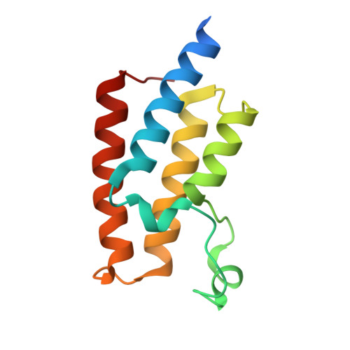
5FE6

Crystal structure of human PCAF bromodomain in complex with fragment ZB1916 (fragment 10)


Experimental Data Snapshot

  • Method: X-RAY DIFFRACTION
  • Resolution: 1.77 Å
  • R-Value Free: 0.212 
  • R-Value Work: 0.179 
  • R-Value Observed: 0.181 

Starting Model: experimental
View more details

wwPDB Validation   3D Report Full Report


Ligand Structure Quality Assessment 


This is version 1.2 of the entry. See complete history


Literature

Structure-Based Identification of Inhibitory Fragments Targeting the p300/CBP-Associated Factor Bromodomain.

Chaikuad, A.Lang, S.Brennan, P.E.Temperini, C.Fedorov, O.Hollander, J.Nachane, R.Abell, C.Muller, S.Siegal, G.Knapp, S.

(2016) J Med Chem 59: 1648-1653

  • DOI: https://doi.org/10.1021/acs.jmedchem.5b01719
  • Primary Citation of Related Structures:  
    5FDZ, 5FE0, 5FE1, 5FE2, 5FE3, 5FE4, 5FE5, 5FE6, 5FE7, 5FE8, 5FE9

  • PubMed Abstract: 

    The P300/CBP-associated factor plays a central role in retroviral infection and cancer development, and the C-terminal bromodomain provides an opportunity for selective targeting. Here, we report several new classes of acetyl-lysine mimetic ligands ranging from mM to low micromolar affinity that were identified using fragment screening approaches. The binding modes of the most attractive fragments were determined using high resolution crystal structures providing chemical starting points and structural models for the development of potent and selective PCAF inhibitors.


  • Organizational Affiliation

    Nuffield Department of Clinical Medicine, Structural Genomics Consortium and Target Discovery Institute, University of Oxford , Old Road Campus Research Building, Roosevelt Drive, Oxford, OX3 7DQ, U.K.


Macromolecules
Find similar proteins by:  (by identity cutoff)  |  3D Structure
Entity ID: 1
MoleculeChains Sequence LengthOrganismDetailsImage
Histone acetyltransferase KAT2B
A, B
119Homo sapiensMutation(s): 0 
Gene Names: KAT2BPCAF
EC: 2.3.1.48 (PDB Primary Data), 2.3.1.57 (UniProt)
UniProt & NIH Common Fund Data Resources
Find proteins for Q92831 (Homo sapiens)
Explore Q92831 
Go to UniProtKB:  Q92831
PHAROS:  Q92831
GTEx:  ENSG00000114166 
Entity Groups  
Sequence Clusters30% Identity50% Identity70% Identity90% Identity95% Identity100% Identity
UniProt GroupQ92831
Sequence Annotations
Expand
  • Reference Sequence
Small Molecules
Ligands 3 Unique
IDChains Name / Formula / InChI Key2D Diagram3D Interactions
5WZ
Query on 5WZ

Download Ideal Coordinates CCD File 
K [auth A](4-azanylpiperidin-1-yl)-cyclopropyl-methanone
C9 H16 N2 O
GCYSQXZPDQBVCH-UHFFFAOYSA-N
DMS
Query on DMS

Download Ideal Coordinates CCD File 
N [auth B]DIMETHYL SULFOXIDE
C2 H6 O S
IAZDPXIOMUYVGZ-UHFFFAOYSA-N
EDO
Query on EDO

Download Ideal Coordinates CCD File 
C [auth A]
D [auth A]
E [auth A]
F [auth A]
G [auth A]
C [auth A],
D [auth A],
E [auth A],
F [auth A],
G [auth A],
H [auth A],
I [auth A],
J [auth A],
L [auth B],
M [auth B]
1,2-ETHANEDIOL
C2 H6 O2
LYCAIKOWRPUZTN-UHFFFAOYSA-N
Experimental Data & Validation

Experimental Data

  • Method: X-RAY DIFFRACTION
  • Resolution: 1.77 Å
  • R-Value Free: 0.212 
  • R-Value Work: 0.179 
  • R-Value Observed: 0.181 
  • Space Group: H 3
Unit Cell:
Length ( Å )Angle ( ˚ )
a = 99.939α = 90
b = 99.939β = 90
c = 99.672γ = 120
Software Package:
Software NamePurpose
REFMACrefinement
XDSdata reduction
SCALAdata scaling
PHASERphasing

Structure Validation

View Full Validation Report


Ligand Structure Quality Assessment 


Entry History 

Deposition Data

Revision History  (Full details and data files)

  • Version 1.0: 2016-01-13
    Type: Initial release
  • Version 1.1: 2016-03-30
    Changes: Database references
  • Version 1.2: 2024-01-10
    Changes: Data collection, Database references, Refinement description