5F4Q | pdb_00005f4q

Crystal structure of the human egg surface protein Juno


Experimental Data Snapshot

  • Method: X-RAY DIFFRACTION
  • Resolution: 1.80 Å
  • R-Value Free: 
    0.227 (Depositor), 0.227 (DCC) 
  • R-Value Work: 
    0.196 (Depositor), 0.191 (DCC) 
  • R-Value Observed: 
    0.199 (Depositor) 

Starting Model: experimental
View more details

wwPDB Validation 3D Report Full Report

Validation slider image for 5F4Q

Ligand Structure Quality Assessment 


This is version 1.5 of the entry. See complete history

Literature

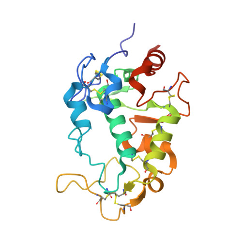
Molecular architecture of the human sperm IZUMO1 and egg JUNO fertilization complex.

Aydin, H.Sultana, A.Li, S.Thavalingam, A.Lee, J.E.

(2016) Nature 534: 562-565

  • DOI: https://doi.org/10.1038/nature18595
  • Primary Citation Related Structures: 
    5F4E, 5F4Q, 5F4T, 5F4V

  • PubMed Abstract: 

    Fertilization is an essential biological process in sexual reproduction and comprises a series of molecular interactions between the sperm and egg. The fusion of the haploid spermatozoon and oocyte is the culminating event in mammalian fertilization, enabling the creation of a new, genetically distinct diploid organism. The merger of two gametes is achieved through a two-step mechanism in which the sperm protein IZUMO1 on the equatorial segment of the acrosome-reacted sperm recognizes its receptor, JUNO, on the egg surface. This recognition is followed by the fusion of the two plasma membranes. IZUMO1 and JUNO proteins are indispensable for fertilization, as constitutive knockdown of either protein results in mice that are healthy but infertile. Despite their central importance in reproductive medicine, the molecular architectures of these proteins and the details of their functional roles in fertilization are not known. Here we present the crystal structures of human IZUMO1 and JUNO in unbound and bound conformations. The human IZUMO1 structure exhibits a distinct boomerang shape and provides structural insights into the IZUMO family of proteins. Human IZUMO1 forms a high-affinity complex with JUNO and undergoes a major conformational change within its N-terminal domain upon binding to the egg-surface receptor. Our results provide insights into the molecular basis of sperm-egg recognition, cross-species fertilization, and the barrier to polyspermy, thereby promising benefits for the rational development of non-hormonal contraceptives and fertility treatments for humans and other mammals.


  • Organizational Affiliation
    • Department of Laboratory Medicine and Pathobiology, Faculty of Medicine, University of Toronto, Toronto, Ontario M5S 1A8, Canada.

Macromolecule Content 

  • Total Structure Weight: 99.61 kDa 
  • Atom Count: 7,594 
  • Modeled Residue Count: 790 
  • Deposited Residue Count: 860 
  • Unique protein chains: 1

Macromolecules

Find similar proteins by:|  3D Structure
Entity ID: 1
MoleculeChains  Sequence LengthOrganismDetailsImage
Sperm-egg fusion protein Juno
A, B, C, D
215Homo sapiensMutation(s): 0 
Gene Names: IZUMO1RFOLR4JUNO
UniProt & NIH Common Fund Data Resources
Find proteins for A6ND01 (Homo sapiens)
Explore A6ND01 
Go to UniProtKB:  A6ND01
PHAROS:  A6ND01
GTEx:  ENSG00000183560 
Entity Groups
Sequence Clusters30% Identity50% Identity70% Identity90% Identity95% Identity100% Identity
UniProt GroupA6ND01
Glycosylation
Glycosylation Sites: 1Go to GlyGen: A6ND01-1
Sequence Annotations
Expand
Reference Sequence

Small Molecules

Ligands 3 Unique
IDChains Name / Formula / InChI Key2D Diagram3D Interactions
NAG

Query on NAG


Download:Ideal Coordinates CCD File
E [auth A],
H [auth B],
J [auth C],
M [auth D]
2-acetamido-2-deoxy-beta-D-glucopyranose
C8 H15 N O6
OVRNDRQMDRJTHS-FMDGEEDCSA-N
GOL

Query on GOL


Download:Ideal Coordinates CCD File
G [auth A],
L [auth C],
O [auth D]
GLYCEROL
C3 H8 O3
PEDCQBHIVMGVHV-UHFFFAOYSA-N
CL

Query on CL


Download:Ideal Coordinates CCD File
F [auth A],
I [auth B],
K [auth C],
N [auth D]
CHLORIDE ION
Cl
VEXZGXHMUGYJMC-UHFFFAOYSA-M

Experimental Data & Validation

Experimental Data

  • Method: X-RAY DIFFRACTION
  • Resolution: 1.80 Å
  • R-Value Free:  0.227 (Depositor), 0.227 (DCC) 
  • R-Value Work:  0.196 (Depositor), 0.191 (DCC) 
  • R-Value Observed: 0.199 (Depositor) 
Space Group: P 41
Unit Cell:
Length ( Å )Angle ( ˚ )
a = 73.15α = 90
b = 73.15β = 90
c = 163.1γ = 90
Software Package:
Software NamePurpose
PHENIXrefinement
Aimlessdata scaling
PDB_EXTRACTdata extraction
XDSdata reduction
PHASERphasing

Structure Validation

View Full Validation Report


Ligand Structure Quality Assessment 


Entry History 

Deposition Data

Revision History  (Full details and data files)

  • Version 1.0: 2016-06-15
    Type: Initial release
  • Version 1.1: 2016-06-29
    Changes: Database references
  • Version 1.2: 2016-07-06
    Changes: Database references
  • Version 1.3: 2020-07-29
    Type: Remediation
    Reason: Carbohydrate remediation
    Changes: Data collection, Database references, Derived calculations, Structure summary
  • Version 1.4: 2023-09-27
    Changes: Data collection, Database references, Refinement description, Structure summary
  • Version 1.5: 2024-11-06
    Changes: Structure summary